As part of the virtual 6th Annual BRAIN Initiative® Investigators Meeting, we hosted the second Show Us Your BRAINs! Photo and Video Contest. In the final article of this series, we feature five of the top entries and the brain cartographers who created them. This year’s contest is open for submissions until May 9 – check it out here.
2nd Place Winner. “Reconstructing the Mind of a Worm”
The C. elegans brain, including every nerve and muscle fiber, reconstructed by serial-section electron microscopy. Credit: Daniel Witvliet, University of Toronto and Harvard University, Cambridge, MA.
Thirty years ago, it took over a decade for scientists to map the brain of the one-millimeter roundworm, C. elegans. This wiring diagram, known as a connectome, was published as the first complete map of neural connections in an animal’s nervous system. Today, improved microscopy and data processing technology make it feasible to map brain wiring in astonishing detail.
This worm connectome is made of about 300 neurons, which connect to the gut, skin, muscles, and sensory organs. Each color represents a different cell type – green cells are muscle fibers and the other colors code for different types of neurons. It was created by Daniel Witvliet, Ph.D., while finishing his degree in Mei Zhen’s lab at University of Toronto. Now, he works in a lab funded by the BRAIN Initiative to mathematically model the brain wiring of C. elegans as they mature from larvae into adults.
Mapping the neural circuits of a tiny worm requires thousands of ultrathin 30-nanometer brain sections. After sectioning the worms, Dr. Witvliet and his colleagues used an electron microscope to image each slice one-by-one. Each twist and turn of every neuron was traced using a computer mouse. An automated software program was used to combine the images into a full connectome.
To study changes in brain wiring across the worm lifespan, researchers repeated this process eight times, during eight different developmental stages. Despite his animation lasting just 30 seconds, this was a difficult and lengthy process, taking nearly a year to complete, says Dr. Witvliet.
Daniel Witvliet (@dwitvliet) received his Ph.D. from the University of Toronto and is now a postdoctoral researcher in Dr. Aravi Samuel’s lab at Harvard University where he studies worm nerve cell wiring and function. He recently developed NemaNode, an online tool to help scientists visualize neural networks.
“Audiovisual Animation of a Gastropod Neural Network”
The neural network of a gastropod seen and heard at the single-neuron level. Each color represents a functional ensemble, a group of neurons with a similar activity pattern. Credit: Jeffrey W. Brown, Evan S. Hill, and William N. Frost, Rosalind Franklin University of Medicine and Science, North Chicago, IL.
Shown here is the neural network of a marine gastropod, commonly known as a sea slug. The video was created by Jeffrey Brown, Ph.D., a postdoctoral research fellow at the Rosalind Franklin University of Medicine and Science. He works with a research team that is part of a BRAIN project aimed at exploring how genes and neural activity interact to guide behavior in the sea slug. They’re also mapping the slug connectome.
To study the neural underpinnings of slug behavior, Dr. Brown and colleagues used a powerful optical imaging system equipped with a photodiode array. This tool monitors the activity of hundreds of neurons simultaneously, nearly 1600 times per second, as slugs generate a stereotyped motor program. Neurons were stained with a voltage-sensitive dye designed to change their levels of light absorption when cells are active. Like a blueprint, researchers overlayed the signals of each neuron onto an image of the slug brain they recorded from (left panel). Each cell was color coded based on its firing pattern.
Listen closely and you can recognize the activity of certain neurons as they fire.
“There’s an undeniable beauty in watching a brain light up and hearing it ‘pop’ as it’s driving actual behavior. This can be appreciated no matter what your scientific background,” says Dr. Brown.
There’s an undeniable beauty in watching a brain light up and hearing it ‘pop’ as it’s driving actual behavior. This can be appreciated no matter what your scientific background – Jeffrey Brown, Rosalind Franklin University of Medicine and Science
Jeffrey Brown earned his Ph.D. from the University of Illinois at Urbana-Champaign, where he was also a research assistant professor and science educator. Currently, he is a postdoctoral research associate in Dr. William Frost’s research group. Learn more about his gastropod network studies by visiting the lab website.
3rd Place Winner. “Fly Through a Fly Brain”
Drosophila (fruit fly) neurons reconstructed by artificial intelligence that uses electron microscope images of a whole fly brain. Credit: Amy Sterling, Sebastian Seung, Mala Murthy, Princeton University, Princeton, NJ.
This video takes us through the brain of another tiny creature, the fruit fly, Drosophila melanogaster. It was created by Amy Sterling, a crowdsourcing specialist and the Executive Director of EyeWire, a game to map the mouse retina, at Princeton University. She collaborates with a research team who recently built a similar game platform called FlyWire to map a full wiring diagram of the fly brain.
“There’s no telling what wonders we will behold in the hidden hundred thousand neuron forest of the fly brain,” says Ms. Sterling.
There’s no telling what wonders we will behold in the hidden hundred thousand neuron forest of the fly brain – Amy Sterling, Princeton University
This dreamy animation of a fly brain was made possible by the latest advances in artificial intelligence (AI). Fly brains were sliced and imaged by electron microscopy. Researchers then used sophisticated AI software to reconstruct each neuron and its connections. But this animation is not perfect – a trained eye can spot small errors in individuals cells, according to Ms. Sterling.
That’s where FlyWire comes in.
FlyWire is a collaborative tool that challenges scientists and the general public to map the connectome of the fruit fly brain. Players are invited to help proofread neuron reconstructions, which are openly available and updated in real-time from the whole fly brain.
By pairing powerful AI with human crowdsourcing, ambitious projects such as FlyWire are quicky transforming the rate of discovery in neuroscience, says Ms. Sterling.
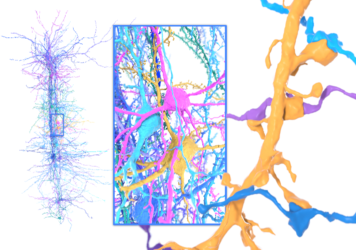
“Column to Synapse”
3D reconstruction of a cortical hypercolumn in the mouse brain. This neural structure was recreated by the Seung lab’s artificial intelligence pipeline. Electron microscope images were collected from a cubic millimeter of the visual cortex. Credit: Amy Sterling, Sebastian Seung, Princeton University, Princeton, NJ.
The fruit fly brain has about 100,000 neurons; the human brain has billions. For now, scientists are working on something in between: mapping the mouse brain. Pictured here is a neural structure called a ‘hypercolumn’ found in the mouse visual cortex.
As larger neural circuits in mice come into focus, neuroscientists hope to better understand how the brain processes information and gain insights into schizophrenia, autism, and other diseases rooted in dysfunctional circuits.
Amy Sterling (@amyneurons) is a crowdsourcing specialist, science communicator, and the Executive Director of EyeWire, a citizen science game that challenges players from around the world to map the mouse brain, one neuron at a time. Check out her TEDTalk to learn more about her creative approach to mapping the mind.
“The Beautiful Brainstem”
23 human brainstem fiber bundles. This brainstem atlas was created using connectome neuroimaging data. Colors represent directionality: top-bottom (blue), left-right (red), front-back (green). Credit: Jim Stanis, Yuchan Tang, Wei Sun, Arthur W. Toga, John M. Ringman, Yonggang Shi, and Ryan Cabeen, USC Mark and Mary Stevens Neuroimaging and Informatics Institute, Los Angeles, CA.
Shown here is an atlas of the human brainstem, the main interface between the brain and body. It was created by Jim Stanis, a scientific visualization artist at the University of Southern California Mark and Mary Stevens Neuroimaging and Informatics Institute. Here, neurobiologists, mathematicians, computer scientists, and animation experts use large-scale neuroimaging datasets, as well as novel software and data analysis tools to build dynamic maps of the brain.
“This is one of the first attempts to comprehensively map the connectomics of the brainstem at this level of detail, charting vastly more descriptive routes of neural highways connecting the brain and body,” says Mr. Stanis.
For this animation, Mr. Stanis and his colleagues used data collected by diffusion MRI, a powerful imaging tool used to map brain structure and connectivity. First, they sifted through an enormous connectome database in search of the cleanest brainstem data. Then they used a 3D modeling technique called tractography to assemble each fiber. Finally, novel mathematical modeling was used to reconstruct 23 fiber bundles for each subject. Data from 20 human subjects was combined to create this colorful brainstem atlas.
With about 100 billion neurons and trillions of neural connections most of the human brain is uncharted. Although we are a long way from mapping the human connectome, advances in brain mapping technology have led to a deeper understanding (and aesthetic appreciation) of the mind.
Jim Stanis is a scientific visualization artist at the USC Mark and Mary Stevens Neuroimaging and Informatics Institute in the Laboratory of Neuro Imaging (@USCLONI). Prior to USC, he received a master’s degree in biomedical visualization and studied fine arts. He also worked as a 3D animator and medical illustrator. Watch more stunning brain animations by Jim and his colleagues here.
The BRAIN Initiative has led to a surge of new technologies to visualize the brain in more ways than ever before. While these innovations have fueled neuroscience research, scientific discovery is driven by the creative individuals who use these tools to uncover the wonders and beauty of the brain, for all to admire.
Feeling inspired? The 2021 contest is open for submissions, check it out here.
Other articles in this series include: