On June 1 and 2, 2020, the 6th Annual BRAIN Initiative® Investigators Meeting was held entirely virtually due to the restrictions posed by the current COVID-19 pandemic. Despite the change to an online platform – run through LabRoots – the conference was a great success! In fact, thousands of investigators, trainees, agency representatives, media, and the public participated this virtual event, many of whom would not have been able to attend if it had been in-person.
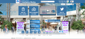
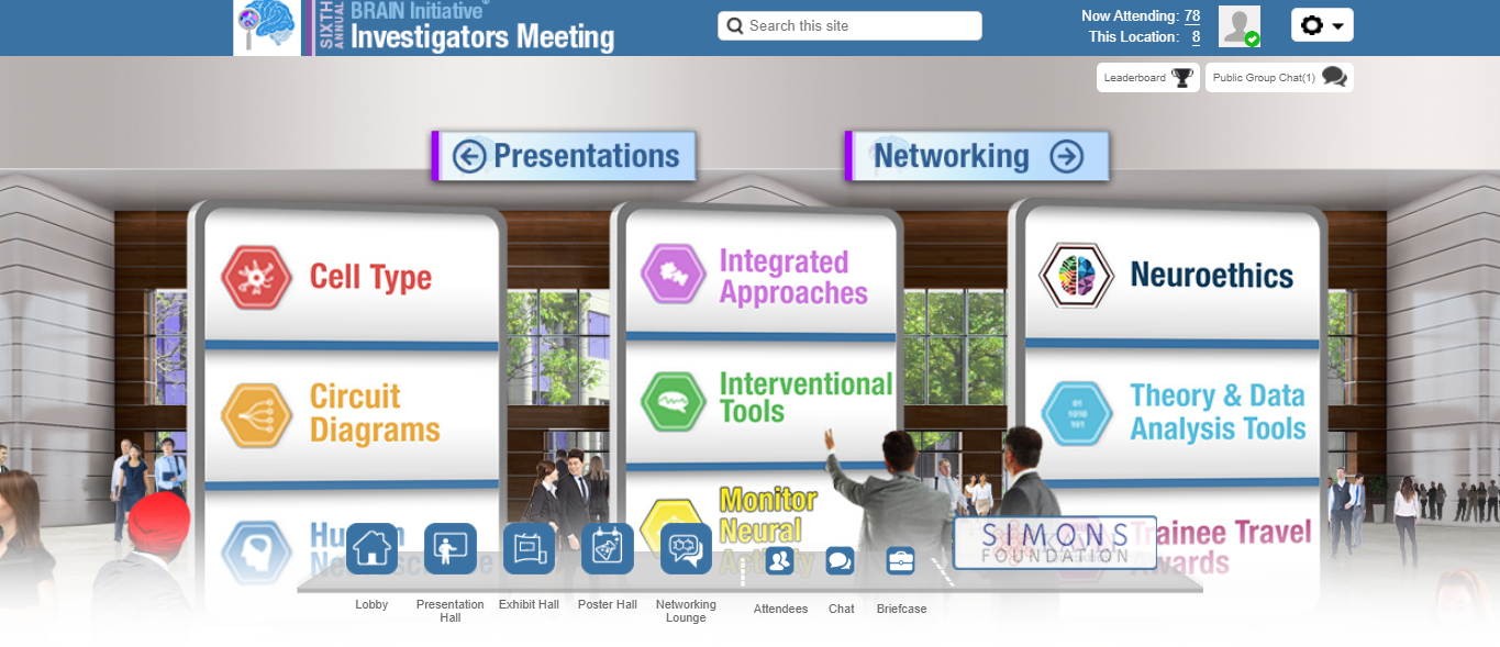
The virtual lobby of the 6th Annual BRAIN Investigators Meeting.
Plenary Keynotes
Each of the plenary keynotes focused on new and exciting breakthroughs made possible by the BRAIN Initiative®
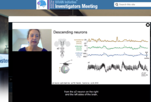
The fist keynote address, on the morning of June 1, was given by Dr. Rachel Wilson from Harvard University. She discussed neural mechanisms of navigation in the fly. Dr. Wilson and her team have taken advantage of new neurotechnologies to map the (female) fly brain to study how navigation behaviors are guided by ‘compass’ neurons. Her lab has even utilized virtual reality in their fly model to answer their questions about receptive fields and microcircuitry of compass neurons.
Dr. Rachel Wilson (Harvard University) gives the first keynote plenary of the 6th Annual BRAIN Investigators Meeting.
Later that day, during the afternoon of June 1, Dr. Eve Marder from Brandeis University gave the second keynote address. During her talk titled “Principles of Circuit Functions from the Study of Small Circuits and Differential Resilience to Perturbations of Circuits with Similar Performance”, Dr. Marder discussed some of the lessons learned from 50 years of studying small circuits in invertebrates. Find out more about her work on small circuits in her recent paper, “Molecular profiling of single neurons of known identity in two ganglia from the crab Cancer borealis,” which can be found here.
Dr. Edward Chang of the University of California, San Francisco, presented the keynote talk on the morning of June 2. He discussed the functional architecture of the speech motor cortex. Speaking is a hallmark of our species, and despite the complexity of speech, we learn it effortlessly as children. Dr. Chang is particularly interested in how the vocal tracts give rise to speech and the importance of vocal pitch in language. The work from his group, along with deep neural network machine learning, may one day enable the reconstruction of words, speech sounds, and vocalizations. This may have important implications for individuals who suffer from brain stem stroke or amyotrophic lateral sclerosis (ALS).
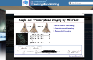
Dr. Xiaowei Zhuang presented the final plenary keynote address on the afternoon of June 2. She discussed single-cell transcriptome imaging through MERFISH, an imaging method that is capable of simultaneously measuring the copy number and spatial distribution of thousands of RNA in a single cell, which was first published in Science in 2015 Dr. Zhuang and her team. Multiple years of hard work were needed to overcome the many challenges that arose when developing this sophisticated imaging technique; made possible by funding from the BRAIN Initiative.
Dr. Xiaowei Zhuang (Harvard University) gives her plenary keynote on the MERFISH technique developed within her lab.
Scientific Symposia
Eight scientific symposia took place during the two-day event and featured many diverse speakers. Viewers were able to engage with all the presenters using a live Q&A session, leading to vibrant discussions. Here we highlight each session – you can view any of them on-demand through the end of May 2021.
Symposium 1: How can dynamical systems neuroscience reciprocally advance machine learning?
Dr. Grace Hwang moderated this session, which featured six speakers who each spoke on some of the challenges and triumphs of machine learning. Dr. Konrad Kording discussed the misleading aspects of current dynamical systems, and Dr. Joseph Monaco asked whether or not transitory neurodynamics can unify learning theories for brains and machine. Next, Dr. Kanaka Rajan discussed neural network models of adaptive and maladaptive state transitions (i.e., active and passive coping) in zebrafish. The session was concluded with discussion of algorithmic dynamics in population codes by Dr. Xaq Pitkow, place cells sequences in hippocampal and prefrontal cortical theta oscillations by Dr. Brad Pfeiffer, and computation in reinforcement learning by Dr. Nathaniel D. Daw.
Symposium 2: Developing and distributing novel electrode technologies
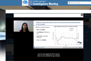
Dr. Allison Yorita (Lawrence Livermore National Laboratory) discusses flexible polymer microelectrode arrays for multi-species applications.
The second session focused on the development and distribution of new electrode array technologies made possible by the BRAIN Initiative. Dr. Cindy Chestek discussed her new high-density carbon fiber microelectrode arrays that are now in beta testing and have demonstrable utility in animal model vagus nerve recordings (see the team’s preliminary report on bioRxiv). Dr. Chong Cie described new ultraflexible neural electrodes that can record from thousands of neurons; these probes overcome the challenges that arise from specialized assembly and implantation of the device. Next, Dr. Tim Hopkins talked about the challenges of design and dissemination, particularly in the context of the next-generation electrodes called Neuropixels probes. Dr. Allison Yorita discussed the fabrication and distribution of flexible polymer electrode arrays and her team’s interest in multi-species applications. Drs. Eric Yttri and Rahul Panat presented the final talk, which described their team’s new, fully customizable, high-density electrode array. This acute 3D nano-printed ‘CMU array’ delivers high-quality neural signals in freely-moving animals.
Symposium 3: Advances in neurotechnologies for human research
Dr. Nanthia Suthana (University of California, Los Angeles) explains her method of recording and stimulating deep brain activity during naturalistic behaviors.
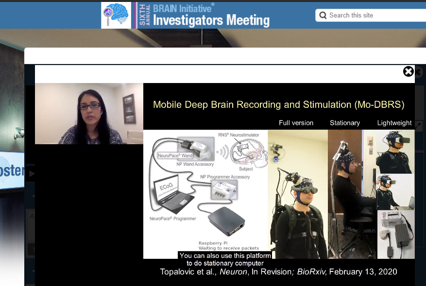
This next symposium focused on what advances in neurotechnology means for human research. First, Dr. Claudi Angeli demonstrated how her team is helping people who have lost their ability to walk to regain their independence using a neurostimulator and computer algorithms. Dr. Nanthia Suthana explained how stimulating and recording deep brain activity could help us understand the neurophysiology of hypervigilance and emotional memory in patients with post-traumatic stress disorder. Dr. Michelle Salas described the Orion Visual Cortical Prosthesis device, which has the goal of artificially enabling vision in blind subjects by directly stimulating the brain. Dr. Harrison Walker talked about how directional (versus circular) deep brain stimulation (DBS) improves gait, side effect thresholds, and therapeutic windows in Parkinson’s disease (PD) patients. Next, Dr. Philip Starr discussed the first instances of recording neural signals from PD patients at home, long term, using his team’s DBS devices. Finally, Dr. Winston Chiong explained the importance of neuroethical considerations when advancing human neurotechnology.
Symposium 4: Expanding species diversity in neuroscience research
This symposium described the variety of species or model organisms in neuroscience research and how that diversity can be expanded upon. The speakers, ranging from trainees to senior scientists, were Drs. Cory Miller, Angeles Salles, Zoe Donaldson, Andrés Bendesky, Paul Katz, and Galit Pelled. These scientists have enhanced our understanding of the nervous system by studying a variety of organisms, including, marmosets, bats, prairie voles, Siamese fighting fish, nudibranchs (mollusks), and octopi. How can studying octopi help us better understand the brain? The number of answers that question has might surprise you. Dr. Pelled’s work with octopi informs our ability to create “smart prosthetic” devices, as discussed here. Each of these species offers unique insights into various niches of neuroscience, from the brain biology of pair bonding (voles) to neural correlates of aggression (Siamese fighting fish).
Symposium 5: Toward petascale and exascale connectomics?
Dr. Jeff Lichtman initiated this session by discussing the history of connectomics. Dr. Alyssa Wilson described how she uses electron microscopy to study mitochondria in the brain to gain insight into circuit development. Dr. Luis Rodriguez explained data and storage challenges, highlighting the volumetric cloud database platform bossDB. Next, graduate student Dannis Cazarez discussed the creation of an open source web portal for neuroscience research that will integrate bossDB and Scalable Analytics for Brain Exploration Research (SABER), a software framework for large-scale imagery data. Ph.D. candidate Jennifer Stiso described her work using neurophysiological data from epilepsy patients to understand how human functional networks respond to perturbations such as seizures. This talk was followed by Dr. Kristen Harris, who discussed developments in transmission-mode scanning electron microscopy (tSEM) tomography for nanoscale resolution. Dr. Eva Dyer described a neural network approach to find patterns in brain region boundaries and her team’s contribution to models of neural architecture through Deep Brain Discovery (DeepBrainDisco!). Finally, Dr. Stephen Plaza discussed building the largest connectomes and the need for “agile” management and robust processes, and Dr. Nuno da Costa closed with a brief talk on cell types and connectomics.
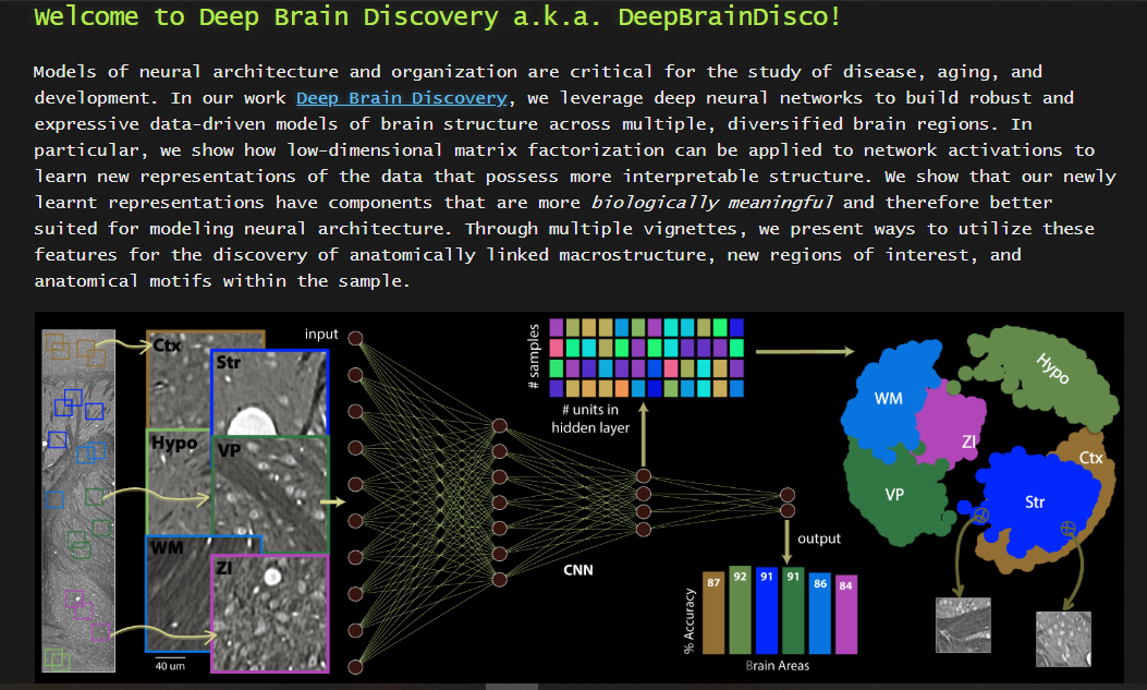
The DeepBrainDisco! webpage was highlighted by Dr. Eva Dyer (Georgia Institute of Technology).
Symposium 6: Advances in human neuroscience
Dr. Helen Mayberg (Icahn School of Medicine at Mount Sinai) aims to reduce symptoms of treatment resistant depression through deep brain stimulation.
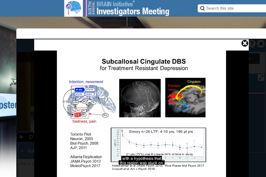
This symposium began with Dr. Jennifer Collinger’s description of a neuroscience-based approach to the restoration of grasp function after spinal cord injury. Then, Dr. Kareem Zhangloul explained the relationship between cortical spiking sequences and memory retrieval in humans. Dr. Helen Mayberg discussed behavioral and brain electrophysiological biomarkers for treatment-resistant depression, while Dr. Chris Abbot described neural correlates of electroconvulsive therapy (ECT) dosing in depression. Finally, Dr. Theodore Satterthwaite spoke on individual variation of association network topography in youth and explained how the topography of the association cortex – the most variable region across individuals – predicts age.
Symposium 7: Molecular taxonomies of brain cells
This symposium focused on recent advances in high-throughput single-cell transcriptomics and epigenomics methods. Specifically, it featured BRAIN Initiative Cell Census Network (BICCN) investigators, whose overarching goal is to generate comprehensive 3D common reference brain cell atlases that will integrate molecular, anatomical, and functional data for describing cell types in mouse, human, and non-human primate brains. Speakers included, Drs. Evan Macosko, Fenna Krienen, Trygvy Baken, Sebestian Preissl, Zhuzhu Zhang, Eran Mikamel, and Bosiljka Tasic. Dr. Macosko’s talk on comprehensive transcriptomics and the cerebellum was particularly interesting. For instance, the cerebellum shows considerable region specialization and has numerous cell types. Dr. Macoskos’s team even identified a new cell type in the molecular layer of the mouse cerebellum. Dr. Krienen also talked on the evolutionary conservation and divergence of brain cell types.
Symposium 8: Emerging high-throughput microscopy methods for imaging brain activity
The final scientific symposium focused on recent developments that allow scientists to record neural activity indicators through larger volumes or with higher framerates than was previously possible. Dr. Valentina Emiliani discussed how optogenetics can be improved so that multiple targets can be manipulated independently in both time and space. Next, graduate student Bowen Wei explained Clear Optically Matched Panoramic Access Channel Technique (COMPACT) for large volume high-throughput deep brain imaging. This was followed by Dr. Sara Abrahamsson, who discussed her team’s 3D imaging using the multi-focus 25-camera miscroscope (M25), for which the open source code in MATLAB is available. Dr. Jerome Mertz described fast multi-plane imaging with reverberation multi-photon microscopy, and Dr. Kasper Podgorski spoke about his work to develop microscopes to track the synaptic activity patterns of single neurons in great detail. Finally, Dr. Na Ji described the advantages of two-photon fluorescent microscopy for in-vivo voltage imaging, and Dr. Aliphasha Vaziri addressed the limitations of diffraction-limited scanning devices and how scanned temporal focusing (s-TeFo) microscopy can be used.
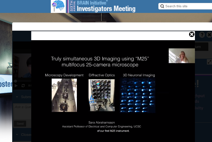
Dr. Sara Abrahamsson (University of California, Santa Cruz) discusses her M25 microscope during the final scientific symposium of the 6th Annual BRAIN Investigators Meeting.
Poster Session and Trainee Spotlights
The virtual poster hall was easily navigated via distinct topics, including neuroethics and the trainee travel awards.
Attendees of the 6th Annual BRAIN Investigators Meeting could also explore a virtual poster hall, where hundreds of posters spanned a variety of topics (see above). This virtual platform enabled users to view and save abstracts, author information, static poster files, as well as listen to pre-recorded audio of poster presenters explaining their work.
This meeting also included two separate Trainee Award Highlight Sessions, where students and postdocs contributing to research funded by the BRAIN Initiative gave flash talks of their research, reinforcing the importance and promise of the trainee workforce. Ph.D. Candidate Nicole Provenza was one of the students highlighted during the Trainee Award Highlight Sessions for her research on how deep brain stimulation can be used to understand and treat obsessive compulsive disorder.
“This month’s meeting was a great opportunity for me to share my work and connect with others in the neuromodulation community. I find these meetings especially useful as a way to stay up to date on new research and ideas.” – Nicole Provenza, Ph.D. Candidate at Brown University
Contests and Interactive Fund
It wasn’t just research at this year’s virtual BRAIN Investigators Meeting. Attendees could earn points to win BRAIN Initiative Alliance sponsored prizes. Points were earned by attending presentations, exploring the exhibit halls, chatting with booth reps, downloading posters, and networking in the virtual lounge. Attendees could also collect extra points by finding the “Swirly Brain” image hidden throughout the event site! Additionally, this meeting featured the second year of the “Show Us Your BRAINS!” Photo & Video Contest, where attendees could vote for each image. Check out the winning entries here!
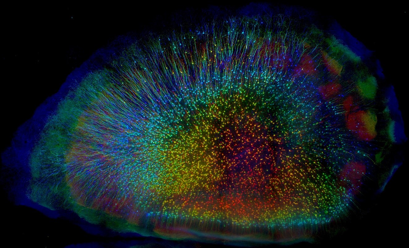
The 1st place winner in the photo contest (entry by Linus Manubens-Gil from Southeast University).
The Communication of Science and Looking Ahead
This BRAIN Initiative® Investigators Meeting also included two science communication workshops, led by experts Liz Neely (The Story Collider) and Rohan Verma (Ogilvy). It is critical that scientists be able to communicate their research not only with other scientists, but also with the public. The two topics covered in this year’s workshops were about 1) using narrative methods and correcting misinformation and 2) how to use social media platforms (such as Twitter) to promote BRAIN Initiative research.
Looking ahead, it is clear that bigger and brighter advances in neuroscience research are in store for the U.S. BRAIN Initiative and its investigators. The virtual platform can be accessed, for free, through the end of May 2021, including all presentations, chat archives from the virtual networking lounge, exhibit booth information, and posters. Although unfortunate circumstances necessitated a virtual event this year, one silver lining might be the engagement of a larger, far-reaching neuroscience community in the efforts made across the BRAIN Initiative.
Please join us at the seventh annual BRAIN Initiative Investigators meeting in 2021!