Need a refresher on the 9th Annual BRAIN Initiative Meeting? Catch up on anything you may have missed here!
Figure 1. The graphic element representing the 9th Annual BRAIN Initiative Meeting.
The 9th Annual meeting of the Brain Research Through Advancing Innovative Neurotechnologies® Initiative, or The BRAIN Initiative®, was held on June 12-13, 2023. For the first year since 2019, the meeting was held in-person in Bethesda, Maryland, and a virtual component was also offered for those unable to attend onsite—the best of both worlds. This year, more than 800 people attended in person, with more than 1,200 people tuning in virtually, some of whom were joining for their first time. These participants came from all over the world, representing investigators, trainees, students, agency representatives, media, and other organizations committed to furthering research in neuroscience. “We’ve had amazing progress in the last year and everyone’s going to hear about it; I’m super excited,” said Dr. John Ngai, Director of the NIH BRAIN Initiative, on the first day of the meeting.
A few key themes stood out at this year’s meeting:
- The role of scientific collaboration was especially present for in-person attendees who could speak with or meet their peers, but it was also a recurring topic in many talks as scientists continue to expand on previous neuroscience discoveries and use each other’s tools for their own research.
- The importance of open science and data sharing was evident—much of today’s research and innovation is made possible thanks to open-source technology, shared datasets, and free access to large data repositories.
- Future neuroethical implications were considered in meeting sessions to remind the audience how to protect neural data and clinical trial participants, especially with the growing body of emerging research that aims to understand the human mind and behavior.
- The promise of artificial intelligence (AI) continued to be a fast-evolving tool for neuroscience development and enhancement, as the audience learned more about how researchers are modernizing their tools with technologies like automation, wearable AI monitors, machine learning, and more.
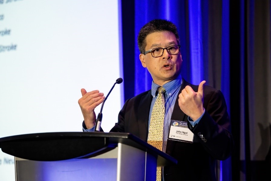
Figure 2. Dr. John Ngai delivering the 9th Annual BRAIN Initiative Meeting opening remarks.
Dr. Ngai opened the meeting by thanking everyone who made the event possible both in-person and virtually. He also encouraged everyone to check out the recently redesigned NIH BRAIN Initiative website and gave a brief overview of the BRAIN Initiative’s mission, goals, and funding history. Dr. Ngai outlined some of the goals of the new BRAIN 2.0 transformative projects, including the BRAIN Initiative Cell Atlas Network (BICAN), BRAIN Initiative Connectivity across Scales (BRAIN CONNECTS), and the Armamentarium for Precision Brain Cell Access. He described the Brain Behavior Quantification and Synchronization (BBQS) program, an effort to develop new platforms and approaches to understand and quantify human behavior. Dr. Ngai also went over the latest training mechanisms that the NIH BRAIN Initiative is applying to enhance diversity, equity, and inclusion in neuroscience.
Show Us Your BRAINs! Photo & Video Contest Winners Announcement
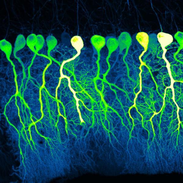
Figure 3. The first-place winner of the Show Us Your BRAINs! Photo Contest, titled, “Dark Commute at 4 a.m.,” by Silas Busch (University of Chicago).
At the end of the opening remarks, Dr. Ngai announced the winners of the fifth annual Show Us Your BRAINs! Photo & Video Contest. Yet again the BRAIN Initiative received many eye-catching and intriguing submissions—check out the winners on the contest’s webpage. Interested in submitting your photo or short video in next year’s contest? Be sure to check the website early next year—all neuroscience researchers are invited to participate regardless of discipline, career stage, or funding source. Congrats to all the finalists for their brilliant contributions!
Plenary Keynotes
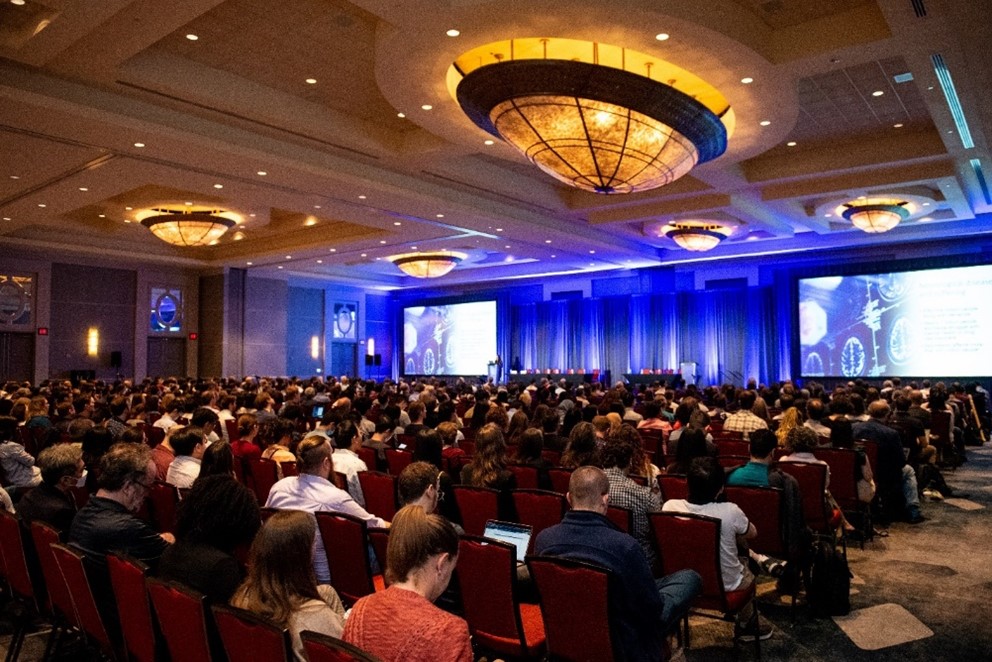
Figure 4. Meeting attendees seated for morning talks on day one of the meeting.
Hundreds tuned in to attend the meeting’s three plenary keynotes, each featuring a notable speaker with fascinating, detailed talks about their research focus areas, how their work was made possible, and the implications their work has for the future of brain science. Each plenary keynote had a Q&A segment to close the session, leading to some fruitful discussions.
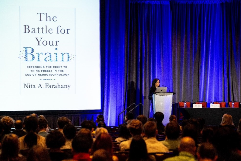
Figure 5. The first plenary speaker, Dr. Nita Farahany, opening her talk titled, “The Battle for Your Brain.”
After the opening remarks, Dr. Ngai introduced the first plenary speaker of day one, Dr. Nita Farahany of Duke University. Her talk, “The Battle for Your Brain,” considered the advantages and ethical implications of using artificial intelligence applications in brain monitoring. Deep brain stimulation and expansive datasets of human neuronal activity can certainly help the neuroscience community learn more about the neurological basis behind conditions like epilepsy or mental health disorders and how to better diagnose and treat them. Consumer neurotechnology is also becoming more mainstream as wearable devices increase in popularity. But with these new applications come with what Dr. Farahany calls a “battle for cognitive liberty,” or the inherent right to our mental privacy and autonomy over our mental experiences. “What if companies could access our thoughts and cognitive processes that shape us? What safeguards should be in place over neural data?” Dr. Farahany questioned. She explained that cognitive liberty is a human right, with many participants in neuroscience research responsible for making sure mental privacy and data is not exploited or commodified. This includes researchers preventing the misuse of data by implementing careful research design practices, commercial entities adopting practices that safeguard our mental privacy and freedom of thought, and investors enabling increased cybersecurity and data management protections.
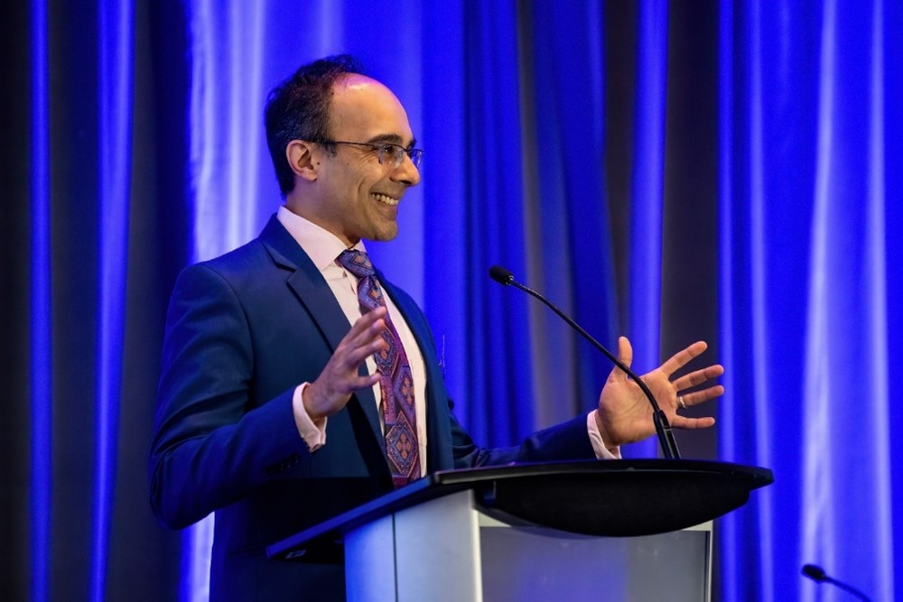
Figure 6. Dr. Sameer Anil Sheth giving his plenary keynote, “Psychiatric Neuromodulation: Pushing the Limits of Personalization.”
Later that afternoon, Dr. Nora Volkow, Director of the National Institute on Drug Abuse (NIDA), introduced the second plenary speaker, Dr. Sameer Anil Sheth (Baylor College of Medicine). His talk, “Psychiatric Neuromodulation: Pushing the Limits of Personalization,” focused on how to treat psychiatric conditions and the push for personalized psychiatric therapy. Dr. Sheth explained how he and his team are studying neuromodulation through implanted devices that can record neural data and how these recordings allow scientists to compare diagnoses, examine activity patterns, and identify biomarkers of treatment response. He provided an overview on the history of stereotactic neurosurgery over the last 50 years and the major research that led to the development of modern-day deep brain stimulation. Dr. Sheth explained that today’s studies investigate conditions like depression and advanced Parkinson’s disease and how therapies affect different features in each disorder. “Are we ready for closed-loop psychiatric neuromodulation?” he asked. While the scientific community supports devices capable of neural recordings, the inter-subject variability is significant, and scientists have much more experience with deep brain stimulation for movement disorders. Dr. Sheth also demonstrated the potential for personalized psychiatric neuromodulation—for example, some psychiatric disorders may benefit from closed loop strategies when symptoms are not constant and have a specific “on” or “off,” such as when tics happen in Tourette’s Syndrome.
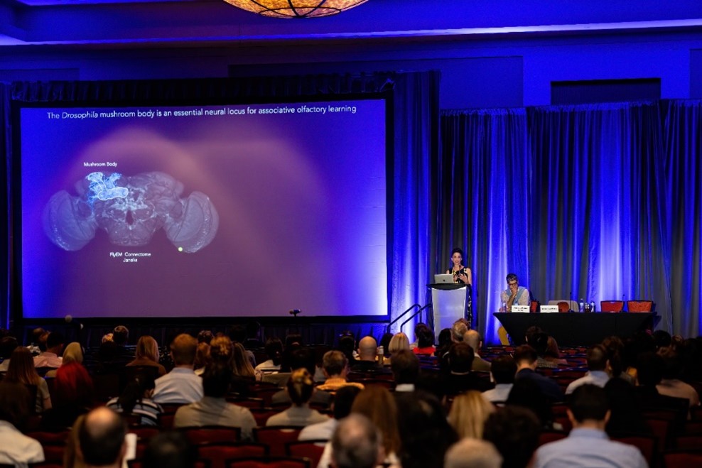
Figure 7. Dr. Vanessa Ruta during her plenary keynote titled, “Circuit Mechanisms for Flexible and Adaptive Behaviors.”
To kick off day two, Dr. Ngai thanked all the speakers from the previous day before introducing the final plenary talk, “Circuit Mechanisms for Flexible and Adaptive Behaviors,” by Dr. Vanessa Ruta of The Rockefeller University. The talk was moderated by Dr. Bob Datta (Harvard University) and demonstrated the strategies Dr. Ruta’s lab uses to study how animals detect and make sense of the complex chemical world to learn behaviors. Using Drosophilia as a model organism, the lab studies the olfactory system through the mushroom body—an olfactory receptor that assists with learning and memory. Specifically, the lab examines the molecular mechanisms for odor detection and neural mechanisms for odor perception. Dr. Ruta explained how dopamine can act locally within mushroom body compartments to change the way odor information is sent from cells to different output neurons. “There’s really dynamic patterns of dopamine release that adhere to the compartmentalized architecture of the mushroom body,” said Dr. Ruta, “these ongoing patterns of dopamine release may serve to shape ongoing behavior.” She also discussed an experiment her lab conducted to examine how neurons send dopaminergic signals while flies travel in olfactory plumes using a closed loop virtual reality system. Flies use edge tracking, a behavior that relies on spatial navigation signals from the central complex to help reencounter dopaminergic signals within an olfactory plume—demonstrating the interplay between the mushroom body and central complex.
Symposia Talks
In addition to the plenary talks, there were six symposia sessions throughout this year’s BRAIN Initiative Meeting. Each session featured a team of panelists who gave a short talk, followed by a Q&A session with the audience.
Symposium 1: Cellular Atlasing and Analysis in Human and Non-human Primate Brain
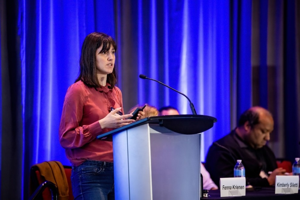
Figure 8. Dr. Kimberly Siletti speaking to the symposium audience.
The first symposia talk of day one was organized by Dr. Trygve Bakken (Allen Institute) and Dr. Edward Lein (Allen Institute) to showcase some of the impactful work being done as part of the BRAIN Initiative Cell Census Network (BICCN). Dr. Fenna Krienen (Princeton University) discussed the need for largescale cellular atlasing efforts in non-human primate brains and the ethical implications involved as these efforts continue to scale up, then presented some research based off a dataset that encompassed 2.4 million brain cells in an adult marmoset brain. Next, Dr. Noah Snyder-Mackler (Arizona State University) gave a virtual talk about a multi-omic atlas of the adult macaque brain. “This talk is taking you on a rapid tour across millions of years of molecular and cellular evolution in the primate brain,” said Dr. Synder-Mackler. His work seeks to understand cell types and pinpoint those that may cause neurological diseases such as attention deficit hyperactivity disorder (ADHD).
Dr. Kimberly Siletti (Karolinska Institutet) discussed an approach to characterize cellular diversity across the human brain based on an atlas with over 100 dissections. She encouraged profiling in every brain region to understand how cell types are composed across regions. Dr. Nelson Johansen (Allen Institute) presented a study that examined how cell types vary in 75 human donors. The study found a few examples of variations that were related to donor demographics and genetics. Dr. Johansen ended his talk by announcing the BICCN Challenge, an initiative beginning in September 2023 where teams can build models to help target cell types. Dr. Timothy Brown closed the session, using his neuroethics expertise to propose solutions for neuroethical issues in the Human Mammalian Brain Atlas program including underrepresentation, recruitment strategies, donor brain criteria, and how data is used, analyzed, and shared.
Symposium 2: The Octopus Brain: Genomics, Connectomics, and Function
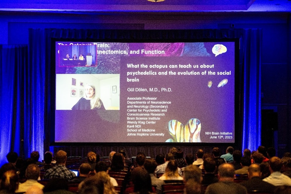
Figure 9. Virtual panelist Dr. Gül Dölan opens her talk while the in-person speakers and attendees watch.
“Who here currently works on octopuses?” said Dr. Paul Katz (University of Massachusetts Amherst), opening another morning symposium session on day one. Nobody in the audience raised their hand. “Who here wishes they worked with the octopus?” he asked. Almost everybody raised their hand. In this session, speakers took turns discussing different facets of octopus research. Dr. Carrie Albertin (University of Chicago) gave insights about sequencing the genome of octopus and squid models to a chromosomal scale. She emphasized the importance of cephalopod biology and studying the octopuses’ developmental mechanisms because of their large nervous systems with vertebrates. Dr. Flavie Bidel (University of Minnesota) demonstrated how she uses volume electron microscopy to map neural connections in the vertical lobe of the octopus brain, showing an in vivo recording during prey capture assay.
Dr. Galit Pellad (Michigan State University) explained how she studies sensory motor circuits and how to image and manipulate them. She showed how her lab is using AI for movement analysis to study how the octopus has great motor control, conducting high-resolution arm movement tracking. Next, Dr. Gül Dölan (Johns Hopkins University) closed the session with a discussion on what the octopus can teach about psychedelics and how the social brain evolves. For example, despite octopuses being asocial and evolutionarily dissimilar to humans, they can exhibit the same behavioral response to 3,4-methylenedioxymethamphetamine (MDMA). For those interested in learning more about octopus neuroscience research, learn more about the Cephalopod Neuroscience Conference coming up in April 2024.
Symposium 3: Functional Manipulation of the Central Nervous System: Theory of the Mind Meets Clinical Intervention
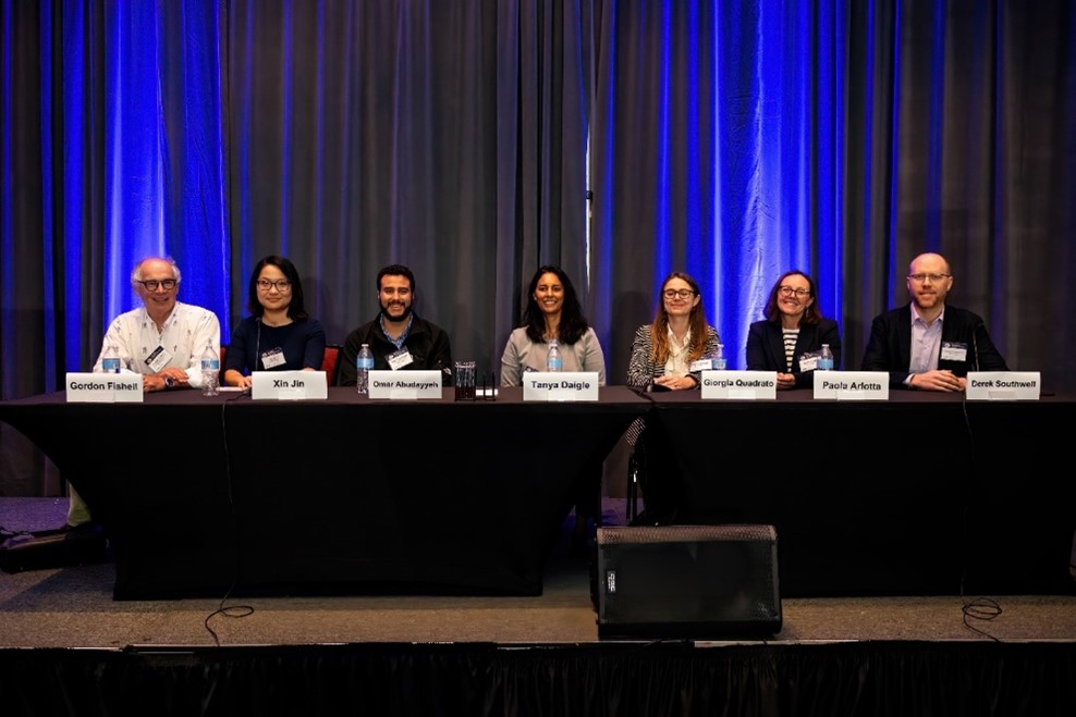
Figure 10. The “Functional Manipulation of the Central Nervous System: Theory of the Mind Meets Clinical Intervention” symposium panel of speakers prior to the start of the session.
Later in the afternoon, Dr. Gordon Fishell (Harvard University) introduced a panel who talked about the methods they use to investigate what genetic factors affect brain cell types. Dr. Xin Jin (Scripps Research) spoke about gene function and the tools her team builds for genetic perturbation, such as Perturb-seq, which uses in vivo perturbation for scalable single-cell transcriptomic analysis in rodents. Dr. Omar Abudayyeh (Massachusetts Institute of Technology) discussed how his lab’s programmable tool, RADARS, or RNA sensing using adenosine deaminases acting on RNA (ADAR), can help with disease modeling in vivo, immune cell regulation, gene editing, and gene therapies by targeting any RNA marker. RADARS can also detect specific mutations in RNA. Dr. Tanya Daigle (Allen Institute) spoke about the Allen Institute’s efforts to discover and use enhancers to target different cell types. She outlined how enhancer adeno-associated viruses (AAVs) are a great method for perturbing cell types within circuits and targeted gene delivery to develop therapeutics.
Next, Dr. Giorgia Quadrato (University of Southern California) discussed the importance of brain development modeling and disease modeling in living human brain tissue. “We have very limited access to human brain tissue,” said Dr. Quadrato, “improvement in human organoids is very important when we think about accelerating clinical translations of these new genetic tools.” To further expand on how some of the tools discussed in the session are being used for clinical translational purposes, Dr. Derek Southwell (Duke University) described an RNA sensing tool called Cell access through RNA sensing by Endogenous ADAR (CellREADR) that was established with Dr. Josh Huang’s lab at Duke University. CellREADR can be used on ex vivo tissue from neurosurgery to monitor and manipulate specific cell types. The session closed with a discussion led by Dr. Southwell and Dr. Paola Arlotta (Harvard University) about how the tools mentioned can be deployed in people.
Symposium 4: Human Neuroscience in the Wild
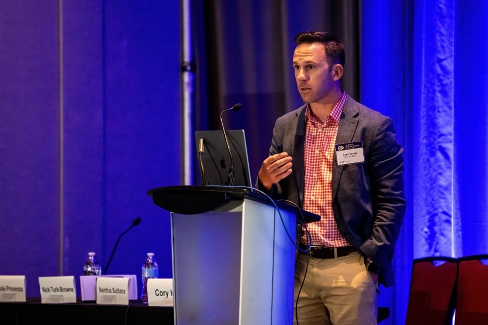
Figure 11. Dr. Cory Inman opening the “Human Neuroscience in the Wild” symposium session.
In parallel, the “Human Neuroscience in the Wild” symposium on day one highlighted advanced human neuromodulation technology applications in the real world, such as mobile eye tracking, mobile movement tracking, and augmented reality. Deep brain recording in freely moving humans is challenging due to the limits of these technologies, but scientists are working to study disorders such as obsessive-compulsive disorder (OCD), epilepsy, and Parkinson’s disease by implanting electrodes in precise brain regions. This session was moderated by speaker Dr. Cory Inman (University of Utah), who gave an overview of his team’s recent work studying memory and event segmentation via hippocampal recordings and eye tracking in humans.
Dr. Nanthia Suthana (University of California, Los Angeles) also showcased examples of eye tracking experiments and how her lab studies memory retrieval using virtual reality navigation and recall. She presented findings demonstrating increases in theta oscillations in the brain whether by successful memory retrieval or internally generated. Dr. Nick Turk-Browne (Yale University) further expanded on human learning and memory by discussing the ways certain aspects of cognition interact with perception, attention, and decision-making. He described some early responsive neurostimulation (RNS) studies and how they conducted human motor control experiments to explore the properties of the hippocampus. Lastly, Dr. Nicole Provenza (Baylor University) talked about her work to help improve the efficacy of deep brain stimulation therapy for treatment-resistant OCD. Patients can use neural recording apps to record at home, while scientists can use that data to help identify neural biomarkers of OCD intensity.
Symposium 5: Sensing, Controlling, and Integrating Brain Processes with Biological Light
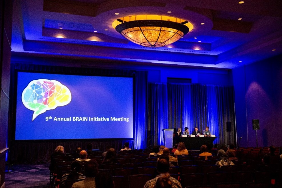
Figure 12. The “Sensing, Controlling, and Integrating Brain Processes with Biological Light” symposium panel during the Q&A portion at the end of the session.
On day two, Dr. Ute Hochgeschwender (Central Michigan University) moderated and presented a symposium session on bioluminescence. She discussed the advantages of biological light in optical imaging, such as integrating transcription, optical synapse and enabling a versatile tool platform for functional dissection of brain circuits. Second, Dr. Michael Lin (Stanford University) talked about fluorescence versus bioluminescence. His lab’s work started with fluorescence, then they made a kinase-modulated bioluminescent indicator called KiMBI to study kinase activity that may contribute to brain cancers and neurodegeneration.
Up next was Dr. Nathan Shaner (University of California, San Diego), who discussed bioluminescent genetically encoded calcium indicators (GECIs). His team developed a system called C-Alpha Based Low-resolution Annotation Method (CaBLAM), a GECI made from an improved luciferase that detects spontaneous and stimulated activity in hippocampal neurons in rats. In the future, the lab hopes to improve its brightness, kinetics, and fusions to optogenetic elements. Dr. Eric Petersen (Central Michigan University) discussed the development and testing of his lab’s BioLumINescent Glutamate indicator (BLING), a bioluminescent neurotransmitter sensor. He explained how glutamate-dependent neuromodulation works, and how his lab makes neurons brighter in rat models by using BLING technology. He closed the session by describing this technology’s applications, such as circuit interrogation and studies in Alzheimer’s disease, Parkinson’s disease, and stroke.
Symposium 6: Reproducible Workflows for Collaborative Neuroscience
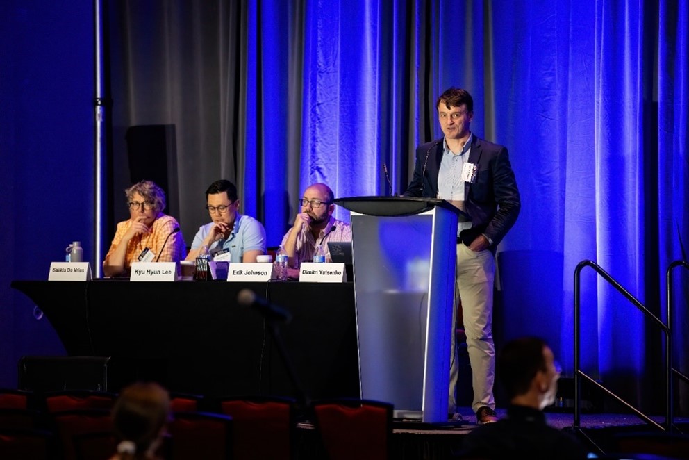
Figure 13. Dr. Dimitri Yaksenko gives his talk as part of the “Reproducible Workflows for Collaborative Neuroscience” symposium session.
Concurrently, Dr. Dimitri Yatsenko (DataJoint) introduced a session about how technologies like automated workflows, advanced computing, and team science help create methodologies that are sharable and reproducible across the neuroscience community. Dr. Erik Johnson (Johns Hopkins University) described BossDB, a cloud-based archive that hosts electron microscopy, x-ray microtomography, and related connectomics datasets. BossDB uses automated deployments with special consideration for data management to enable fast data access and sharing. Next, Dr. Kyu Hyun Lee (University of California, San Francisco) spoke about Spyglass, an open-source software framework for neuroscience data analysis. Spyglass comes with automated workflows and is interoperable with the Neurodata Without Borders format to support data sharing and reuse. Dr. Julia Huntenburg (International Brain Laboratory) detailed the International Brain Laboratory, a team science group consisting of 22 labs in 6 countries that studies neural circuitry related to decision-making and behavior.
Dr. Yatsenko returned to give his talk on DataJoint Elements, an open-source framework that supports shared data pipelines. “Our goal is to take what we learn from multi-lab, complex projects and help individual smaller labs implement the same types of processes for efficiency and reproducible workflows in their labs,” said Dr. Yatsenko. Last, Dr. Saskia de Vries (Allen Institute) presented on collaborative neuroscience at the Allen Institute for Neural Dynamics, a new, sister institute to the Allen Institute for Brain Science that focuses exclusively on the neural signals behind complex behaviors. The Institute uses team science with a focus on open resources, accessibility, maintainability, reproducibility, and data exchange.
Poster Sessions and Exhibits
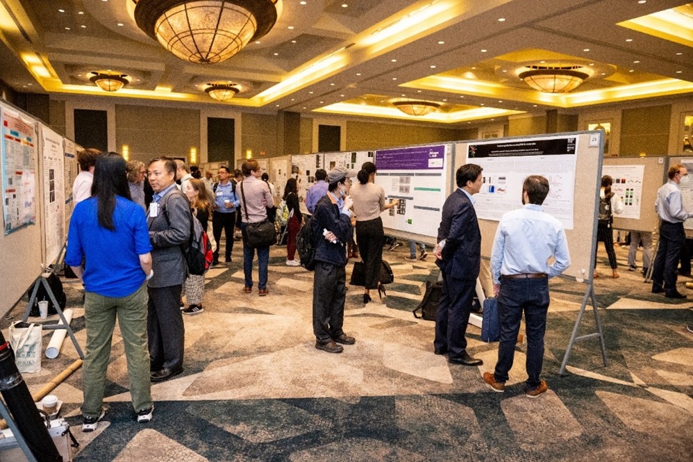
Figure 14. The 9th Annual BRAIN Initiative Meeting poster hall.
There were three dedicated poster sessions at the 9th Annual BRAIN Initiative Meeting. A large poster hall had different presenters at each session for in-person attendees, and virtual attendees could access a gallery where they could chat with presenters and leave questions about posters. As part of the virtual gallery, select posters had pre-recorded audio presentations from the research team with more details about their work. All posters are still available on the meeting website, are organized by topic area, and can be easily downloaded.
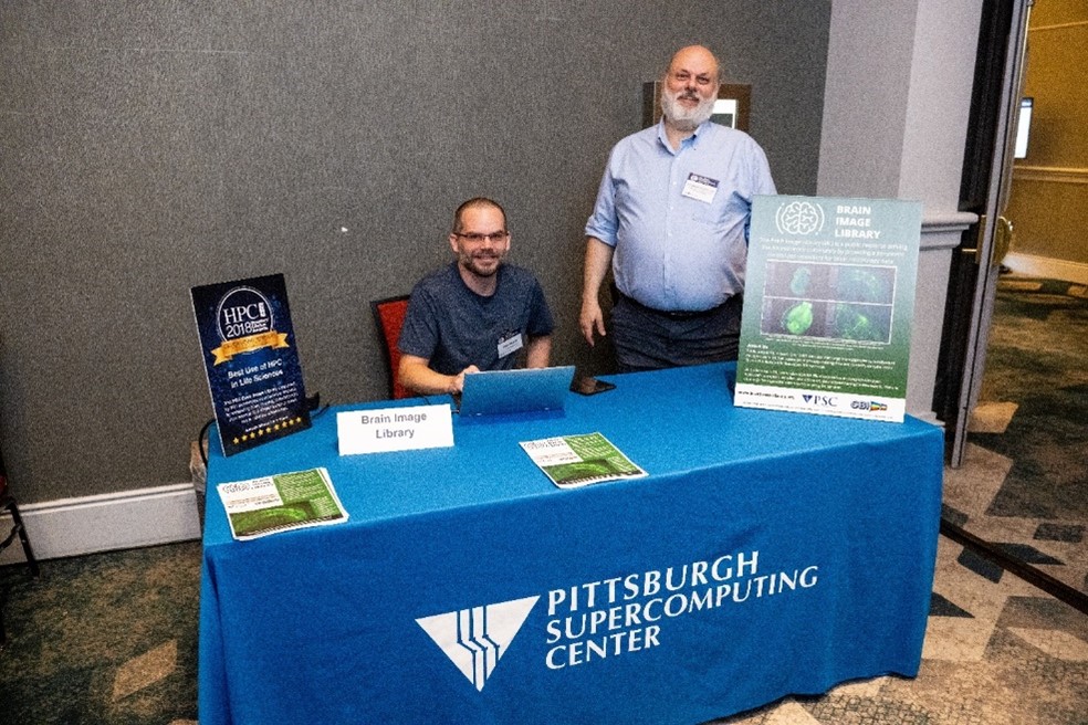
Figure 15. Dr. Alan Watson and Alexander Ropelewski at the Brain Image Library exhibitor table.
Several exhibitors set up booths along the halls at the meeting, and virtual attendees could chat with exhibitors during a special exhibit session at the end of day one. “We have an exhibit table here and a poster, which one of our curators was presenting this morning,” said Dr. Alan Watson, a principal investigator with the University of Pittsburgh who worked the exhibit table for the Brain Image Library. Also manning the Brain Image Library booth was Alexander Ropelewski, a principal investigator at the Pittsburgh Supercomputing Center. “My favorite thing about attending in-person is meeting people to discuss projects, collaborations, or issues that they’re having to see if we can resolve things or give someone a different approach or way of looking at things,” Ropelewski said.
Trainee Highlight Awards
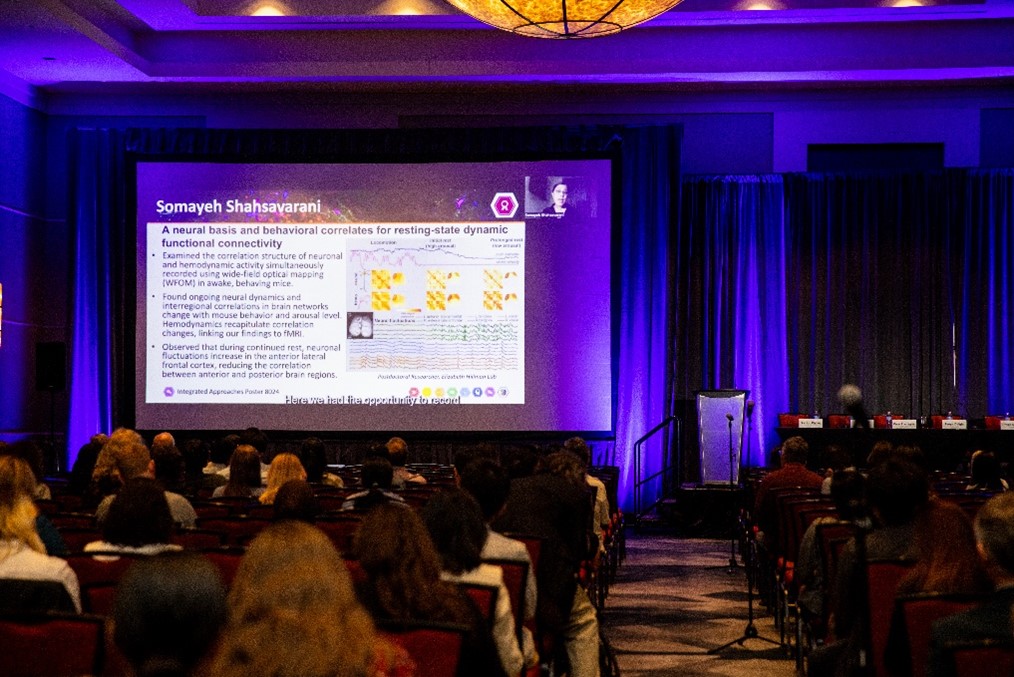
Figure 16. Meeting attendees watching the Trainee Highlight Award presentations.
Each year, the Trainee Highlight Awards showcase the work of early career researchers who might be in high school, undergraduate or graduate programs, medical school, or be postdoctoral fellows or residents. The awards were divided into one session per day during this year’s meeting. During these sessions, 30 trainee highlight awardees gave a three-minute virtual “flash” talk on their research project alongside a scientific poster that described the highlights of their impactful work in neuroscience. Trainees were later available to network and discuss their projects further during the meeting’s poster sessions. For a full list of the awardees and their poster titles, visit the meeting agenda.
Focused Topic (Specialty) Sessions
BRAIN, Neuroscience, and Beyond: Building Our Early Career Community
A special networking event took place the evening before the meeting formally began. Trainees and early career investigators who were attending in-person were invited to socialize and learn more about funding opportunities from their peers. “Meeting new people in-person was so much easier—the early career event was so great,” said Dr. Sumaira Zammurad, a postdoctoral fellow at Columbia University.
A First Look at the Allen Brain Cell Atlas: Multimodal Visual Analysis at Whole-Brain Scale
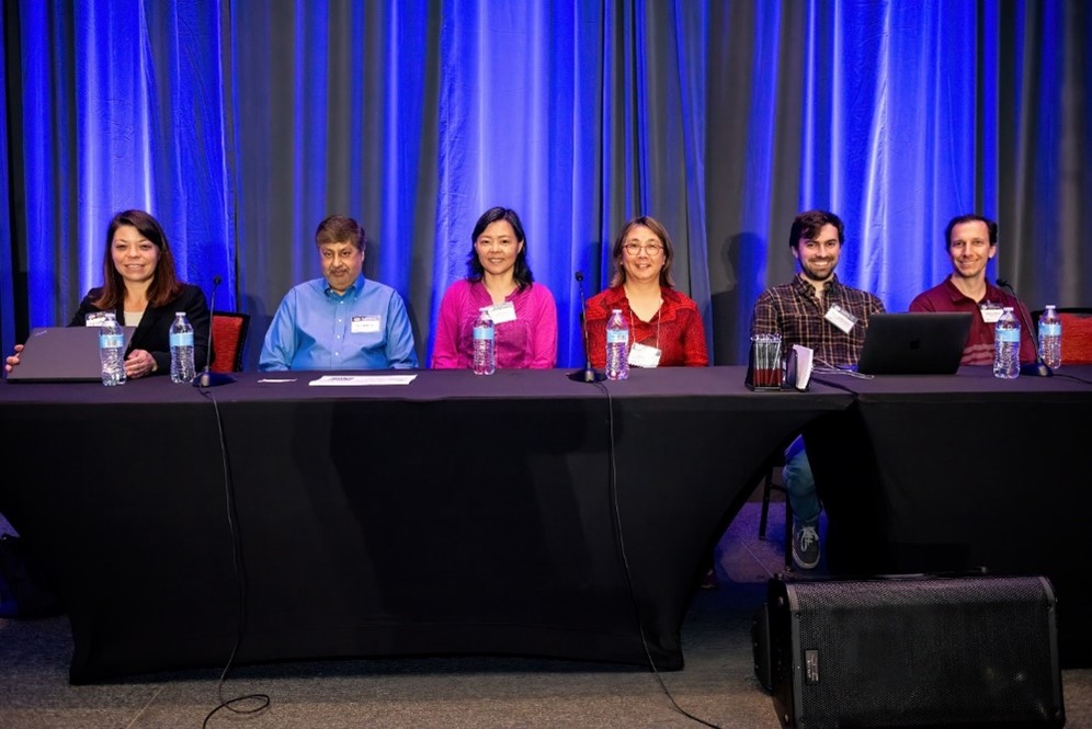
Figure 17. A group of panelists from the Allen Institute.
In this workshop, speakers from the Allen Institute for Brain Science provided an overview and demonstration of the Allen Brain Atlas, a web data portal that serves as a genome-wide map of the adult mouse brain. Speakers also discussed how the Allen Brain Atlas can support the BICCN and the BICAN datasets.
Data Science Consortium Workshop
This workshop featured three speakers, Dr. Manuel Schottdorf (Princeton Neuroscience Institute), Dr. Guoqiang Yu (Virginia Tech), and Dr. Edgar Walker (University of Washington). Each speaker discussed how to improve data science within the neuroscience community, especially within the NIH BRAIN Initiative U19 Data Science Consortium.
National Science Foundation Program on Integrative Strategies for Understanding Neural and Cognitive Systems
Program directors from the National Science Foundation presented research involving Neural and Cognitive Systems (NCS) during this in-person session. Presentations focused on two themes: brain activity in ecologically valid, natural, wild, and social settings, and sensory and motor neuroprosthetics.
Technology and Resource Dissemination: Best Practices and Lessons Learned
Four panelists lent their knowledge on resource dissemination during this in-person session, followed by a networking session. Panelists included Dr. Elizabeth Hillman (Columbia University), Dr. Melina Fan (Addgene), Dr. Megan Peters (University of California, Irvine), and Amy Sterling (Eyewire, Princeton University).
Introduction to the NIH and BRAIN Ecosystem for Translation
In this session, participants heard from investigators who had previously transitioned their own projects to NIH grants and funding. Speakers told stories about their funding journeys, how they overcame challenges, how they budgeted, and how they optimized their projects using NIH technology. Panelists included Dr. Philip Starr (University of California, San Francisco), Dr. Vanessa Tolosa (Meta), and Dr. Jonathan Viventi (Duke University).
Public Engagement Models for the Benefit of BRAIN Initiative Science
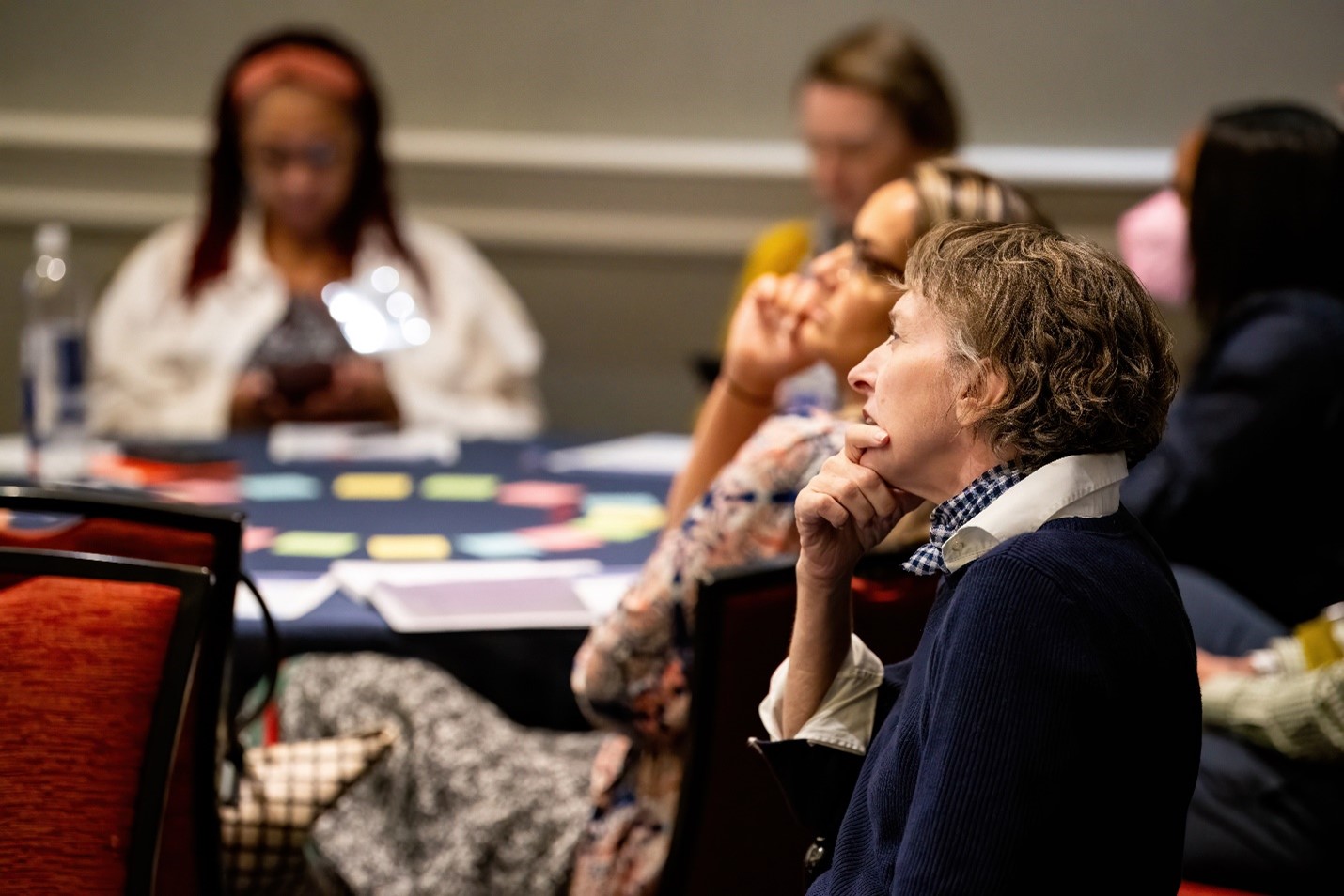
Figure 18. Attendees participated in interactive activities during the “Public Engagement Models for the Benefit of BRAIN Initiative Science” specialty session.
Attendees participated in interactive activities that helped everyone better understand the role of collaboration in public engagement approaches during this in-person session followed by a moderated discussion led by Dr. Caroline Montojo (Dana Foundation) and Dr. Amy Bany Adams (NIH/NINDS). Panelists included Dr. Jayatri Das (Franklin Institute), Dr. Lomax Boyd (Johns Hopkins Berman Institute for Bioethics), Dr. Jesse Isaacman-Beck (NIH), and Layne Oliff (member of NIH/NINDS ALS Strategic Planning Committee).
The Theater of Thought Abridged Documentary Screening and Q&A
The BRAIN Initiative meeting also held a screening for “Theatre of Thought”, a 2022 documentary by Dr. Werner Herzog about advances in neurotechnology and what potential helpful or harmful implications these technologies have in the future.
Meet the Funders
Representatives from the NIH BRAIN Initiative, the Simons Foundation, the Kavli Foundation, and the National Science Foundation participated in this in-person networking session, chatting with attendees to help them learn about funding opportunities.
Conclusion
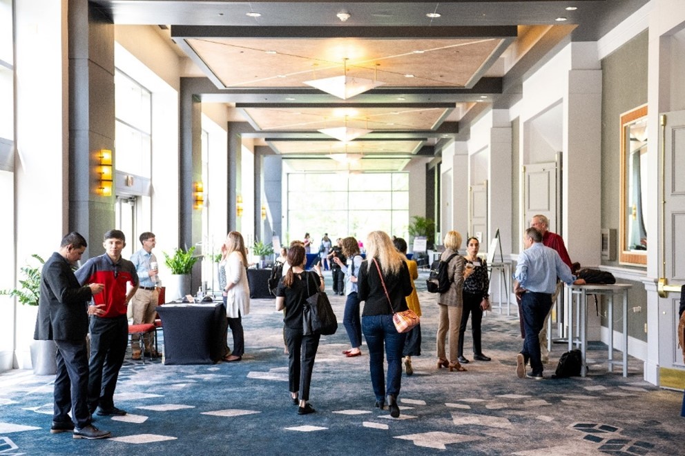
Figure 19. Attendees gathered in the hallway at the meeting.
The 9th Annual BRAIN Initiative Meeting was a tremendous success! Attendees were especially grateful to be back in-person, which sparked new connections and networking opportunities. “I want to show people what we are doing and communicate to find and meet potential collaborators,” said Dr. Jing Xiang (Cincinnati Children’s Hospital), a first-time meeting attendee. Others were excited to learn more about what projects and topics their peers are investigating. “I’m still in the early stages of figuring out my own project and where to take my own work,” said another first-time attendee, Katherine Kabotyanski (Baylor College of Medicine). “Getting a sense of all the newest and coolest things that are being done in this field has been really helpful.”
The learning and collaboration doesn’t need to stop here—revisit any talk or session from this year’s meeting by checking out the on-demand recordings on the meeting website. We hope to see you next year at the 10th Annual BRAIN Initiative Meeting from June 17-18, 2024!
All images provided by Lesnick Photo.