Whether you attended and are looking for a recap or you couldn’t make it and want an overview, we have all the details of the 10th Annual BRAIN Initiative Conference here!
Figure 1. The graphic element representing the 10th Annual BRAIN Initiative Conference.
On June 17–18, 2024, we held the 10th Annual BRAIN Initiative Conference! The meeting was a great success, with more than 800 in-person attendees and 1,700 people virtually tuning in.
To open the conference, Dr. John Ngai, director of the National Institutes of Health (NIH) Brain Research Through Advancing Innovative Neurotechnologies® Initiative, or The BRAIN Initiative®, welcomed attendees and showed a video message from former NIH Director Dr. Monica Bertagnolli, who expressed how proud she is of the vast expertise and innovative tools that have encompassed the NIH BRAIN Initiative over the last decade. Dr. Bertagnolli and Dr. Ngai both spoke on the scope of the BRAIN Initiative, which has supported more than 1,500 projects and thousands of principal investigators, with funding totaling more than $3.5 billion across 10 NIH Institutes and Centers and federal and non-federal partners. Over the last decade, BRAIN Initiative projects have made incredible progress in applying new tools and methods to treat neurological disorders like stroke, chronic pain, Alzheimer’s disease, and obsessive-compulsive disorder (OCD).
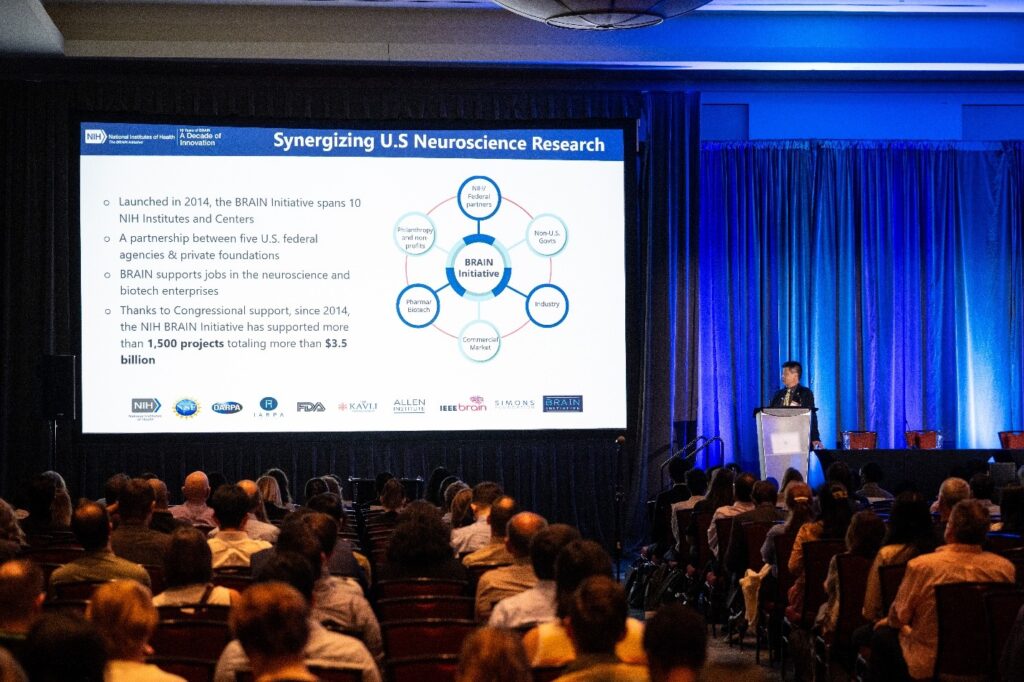
Figure 2. Dr. Ngai opening the 10th Annual BRAIN Initiative Conference.
Dr. Ngai reviewed a few key themes of the BRAIN Initiative in his introduction, all related to keeping neuroscience innovative, open, and ethical. Dr. Ngai stressed the importance of neuroethical implications behind the work that the BRAIN Initiative supports and how the BRAIN Initiative conducts proactive and continuous assessments of neuroethics in all its findings and projects.
“[There are] so many impacts that have come out of the conference in 10 years,” says Dr. Ngai. “Huge resources like our BRAIN [Initiative] Cell Atlas Network [BICAN] are really transforming the way we can do things in the future. What really represents progress and is so impressive is that when the Cell Census [BICCN] project started out in 2014, we did not have the tools to do what’s being done today. It’s been amazing progress both from a technology and science point of view.”
Building on a Decade of Innovation
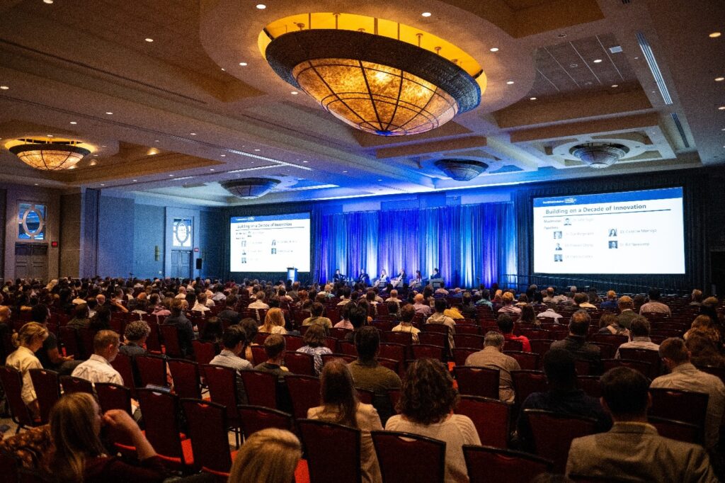
Figure 3. The crowd listening to the Building on a Decade of Innovation session.
On the first day of the conference, we celebrated 10 years of BRAIN Initiative innovation by hosting a panel about our most notable achievements and thinking about what the future may hold in the next decade. Dr. Ngai moderated the Building on a Decade of Innovation session in a fireside chat–style conversation with speakers Dr. Cori Bargmann (Rockefeller University), Dr. Edward Chang (University of California, San Francisco), Dr. Francis Collins (former NIH director), Dr. Caroline Montojo (Dana Foundation), and Dr. Bill Newsome (Stanford University). Dr. Ngai told the story of how the BRAIN Initiative began, showing a video with Dr. Collins and former President Barack Obama first announcing the BRAIN Initiative. “What stuck with me was President Obama’s call to action, which was ‘Let’s get to work,’” said Dr. Ngai. The panelists built on the origin story of the BRAIN Initiative, crediting the Kavli Foundation for “stirring up ideas” about launching a large funding source for brain research and NIH for building it “as a labor of love.” Philanthropy played a large role in the BRAIN Initiative taking off, thanks to many parties contributing ideas, finding ways to address investigator needs, and forming the BRAIN Initiative Alliance. “We thought this was essential,” said Dr. Chang.
The panel also discussed the importance of incorporating ethics into neuroscience research and the early conversations that led to the neuroethical standards we have today. The panelists agreed that neuroscience is loaded with social and policy issues and examined the levels of support and community engagement necessary to keep neuroscience ethical. Since it began, the BRAIN Initiative has made many researchers, doctors, and policymakers think more about the ethical implications of advanced human neuroscience and form new standards and frameworks to protect patients from risks and consent issues.
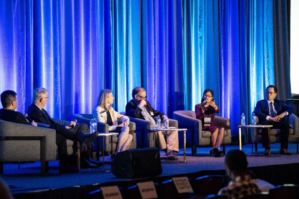
Figure 4. The speakers during the Building on a Decade of Innovation session.
The panel closed by examining the future of the BRAIN Initiative and brain science. Many speakers were surprised by the progress that we’ve made with intervention strategies for brain disorders. “Ten years ago, we were really impressed by how well deep brain stimulation worked for things like Parkinson’s disease…but we’re still just scratching the surface and the potential for really meaningful human interventions is closer than we think,” said Dr. Bargmann. Although the tools we have now are much more powerful than the ones we had just 5 years ago, artificial intelligence (AI) may help us find better ways to compute and understand data. “I think we need to understand model systems better. That’s what I’m super excited about in the coming decade,” said Dr. Chang. The panel continued discussing the trajectories of natural and artificial intelligence and how they will complement each other, considering how we can train the next generation to consider ethical and societal implications to answer the role of science in the world.
Scholar Spotlight Awards
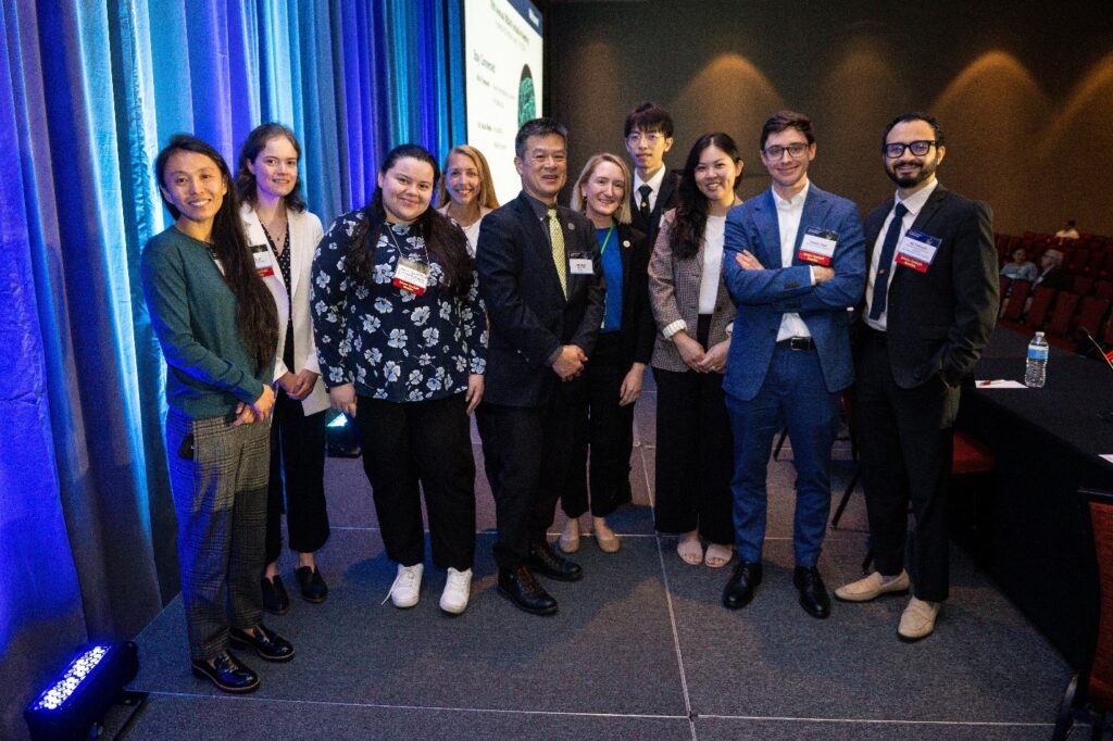
Figure 5. Dr. Ngai with the Scholar Spotlight awardees.
Just after the opening remarks on day one, Dr. Ngai introduced the eight Scholar Spotlight awardees and welcomed them onstage to give a 1-minute “lightning” talk to the crowd to highlight their research objectives, accomplishments, and goals. Awardees spoke about machine learning for stroke rehab, neurostimulation for epilepsy treatment, deep brain stimulation to help OCD, behavioral responses to pain, and more. “Sharing our research is so important. The more I talk about it, the more it’s going to help me and others understand it,” said Scholar Spotlight awardee Dr. Melissa Franch (Baylor College of Medicine). “What’s exciting about the BRAIN Initiative is the willingness to take risks on innovative, high-risk, high-reward research…I like the different areas of the research that the conference brings, and I feel like sharing your work and getting people’s perspectives allows you to get helpful feedback and ideas for your projects.” Learn more about the awardees and their science by viewing the Scholar Spotlights Lightning Talks.
The BRAIN Initiative Photo and Video Contest Winners Announcement
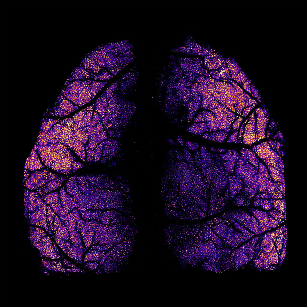
Figure 6. The first-place winner of the The BRAIN Initiative PhotoContest, titled “Whole-cortex scale in vivo two-photon imaging with single-cell resolution in mice,” by Zongyue Cheng (Purdue University).
Each year at the conference, we look forward to announcing the winners of the annual BRAIN Initiative Photo and Video Contest. This year, Dr. Ngai opened day two of the conference by revealing the winners and showcasing their entries, with a special “BRAIN at 10” category to celebrate 10 years of BRAIN-funded research. You can check out all of the finalists on the contest web page. A huge congratulations to our finalists for capturing the creative spirit of the BRAIN Initiative!
Plenary Keynotes
Each year, the plenary keynotes attract a large crowd interested in learning about the next groundbreaking installments in neuroscience. This year, we were grateful to hear from two distinguished plenary speakers, Dr. Liqun Luo of the Howard Hughes Medical Institute and Stanford University, and Dr. Viviana Gradinaru of the California Institute of Technology. A Q&A session closed out each plenary keynote, during which participants were invited up to the mic, or through the virtual chat feature, to ask the speakers questions about their research.
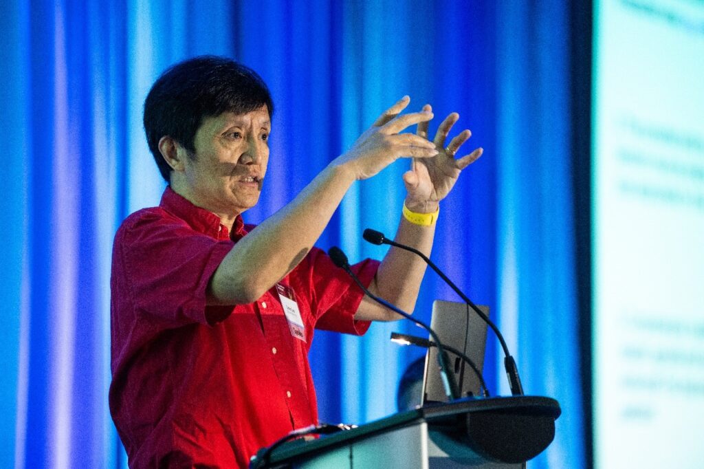
Figure 7. Dr. Liqun Luo presents his plenary talk, “Deconstructing the Serotonin System in the Mouse Brain.”
To kick off the plenary keynotes, Dr. Luo delivered his talk, “Deconstructing the Serotonin System in the Mouse Brain.” The talk overviewed the tools Dr. Luo’s lab develops and uses to map neural circuit function in mammals, such as the TRIO (Tracing the Relationship between Input and Output) and cTRIO (cell-type-specific TRIO) methods. By applying cTRIO, Dr. Luo’s lab found that systems have distinct architectures and that the frontal cortex and central amygdala that project serotonin neurons have two distinct subsystems.
Dr. Luo also walked the audience through three unpublished studies. The first, “Architecture of serotonergic projectome in the entire mouse brain,” showed that serotonin neuron project patterns fall into five projectomic clusters. Some clusters comprise brain regions in the same circuit with similar functions, whereas others have unexpected links and dissociations between brain regions. The second, “Modulation of female social behavior by projection-specific serotonin neurons,” activated serotonin neurons in female mice to find patterns in how they behaved toward mice pups. The study looked at whole-brain axon projection patterns to study pup interactions. The last study, “Deconstructing the serotonin system via the serotonin-receptor axis,” investigated the effects of drugs on spontaneous behavior using motion sequencing and c-Fos mapping. This study enabled joint neurobehavioral modeling and prediction.
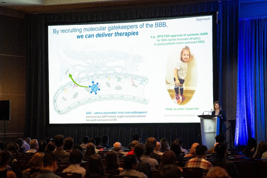
Figure 8. Dr. Viviana Gradinaru during her plenary keynote, “Blood–Brain Barrier: Friend and Foe.”
On day two, Dr. Gradinaru gave her talk, “Blood–Brain Barrier: Friend and Foe,” which described technologies that perturb and monitor the brain, touching on the limits of applying these interventions in larger models. With 1 billion people around the world affected by brain diseases, Dr. Gradinaru emphasized how important it is that we investigate brain-wide access to deliver genetic tools and therapies that help restore brain function. One challenge to delivering those tools or therapies is the blood–brain barrier (BBB), which regulates arrivals to the brain and blocks pathogens so that viruses or bacteria cannot gain entry. But if we can recruit the molecular gatekeepers that guard the BBB and figure out how to create tools and receptors for selective entry, we can deliver therapies.
Dr. Gradinaru’s lab has been bioengineering intravascular adeno-associated virus (AAV) vectors to cross the BBB and enter brain cell types. These AAV capsids have the potential to evolve for specific features and to reach targeted brain areas. The lab built a toolkit of systemic AAVs for various systems from mice to macaques to human cells. Independent researchers are using these tools, and Dr. Gradinaru says her favorite application was for brain-wide genetic screens. Her lab applies them to study Friedreich’s ataxia (FA), a rare neurodegenerative disease. After applying AAV gene therapy in mice, their motor control improved during motor coordination testing. The lab also tested AAV gene therapy to cross the BBB in larger mammalian brains, learning more about basic BBB biology in the process. A major goal of Dr. Gradinaru’s is to understand the mechanisms that underly BBB transport across species and within brains. Dr. Gradinaru discussed approaches to achieve that goal, including how her research led to an unexpected discovery: an ancient conserved enzyme (carbonic anhydrase IV) that can mediate transport across BBB. She closed her talk by iterating the benefits of unlocking and locking blood–brain portals in different evolutionary contexts, such as delivering brain therapeutics to specific cells or regions, navigating health threats, and helping people become more resilient when facing disease.
Symposia Talks
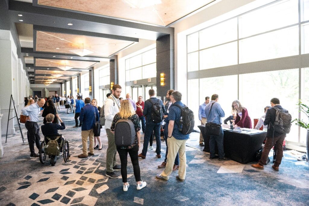
Figure 9. Attendees gathered in the main hallway before the first symposia talks.
The six symposia sessions allowed conference attendees the opportunity to choose the topics most intriguing to them, to hear from expert panelists, and to ask questions at the end of each session. “My favorite part of the conference is the interesting symposiums. There’s always a chance I’ll find inspiration, new methods, or just understand more where the field is at by attending them,” said conference attendee Dr. Gene Yu (Duke University). The symposium speakers were just as excited as the attendees to share their work. Some of the symposia sessions ran concurrently, so if you missed one you can catch up by signing in to the conference platform with your conference credentials or watch on YouTube and select the 2024 BRAIN Conference playlist.
Symposium 1: High-density and High-resolution Neurophysiology to Reveal Local Microcircuits in the Human Brain
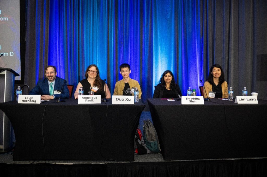
Figure 10. The panelists from the High-density and High-resolution Neurophysiology to Reveal Local Microcircuits in the Human Brain session.
This session, moderated by Dr. Leigh Hochberg (Brown University), highlighted approaches for high-density and high-resolution neurophysiology, including therapy delivery and neuroethical implications. Dr. Angelique Paulk (Massachusetts General Hospital/Harvard University) discussed technological advances in high-resolution brain recording devices. High-resolution recordings increase our understanding of cortical layer physiology and how brain circuits affect cognition. For example, when used in the operating room, Neuropixels can capture single cell activity in and around cancerous tumors. The second speaker, Dr. Duo (Many) Xu (University of California, San Francisco), presented on the speech production by single neurons. His work uses high-resolution recordings to find patterns that investigate how “mirror” and “bridge” neurons in human precentral gyrus connect speech content from perception to production.
Next, Dr. Shraddha Shah (Baylor College of Medicine) spoke about how her lab uses Neuropixels and audio cues for high-density and large-scale recordings to test functional responses in human hippocampus single neurons, testing patient-specific stimuli and functional responses under anesthesia to find out the functional and physiological properties of human single neurons in a temporal cortical microcircuit. Last, Dr. Lan Luan (Rice University) presented on electrode development and engineering for animal research. Her lab created nanoelectronic threads (NET), or bi-directional, long-lasting, ultraflexible electrodes that are stable and scalable. These electrodes record and track neurons for unit activity and connectivity mapping.
Symposium 2: Imaging Outside-the-Toolbox: Novel Solutions for Large Scale and Deep-Tissue Imaging
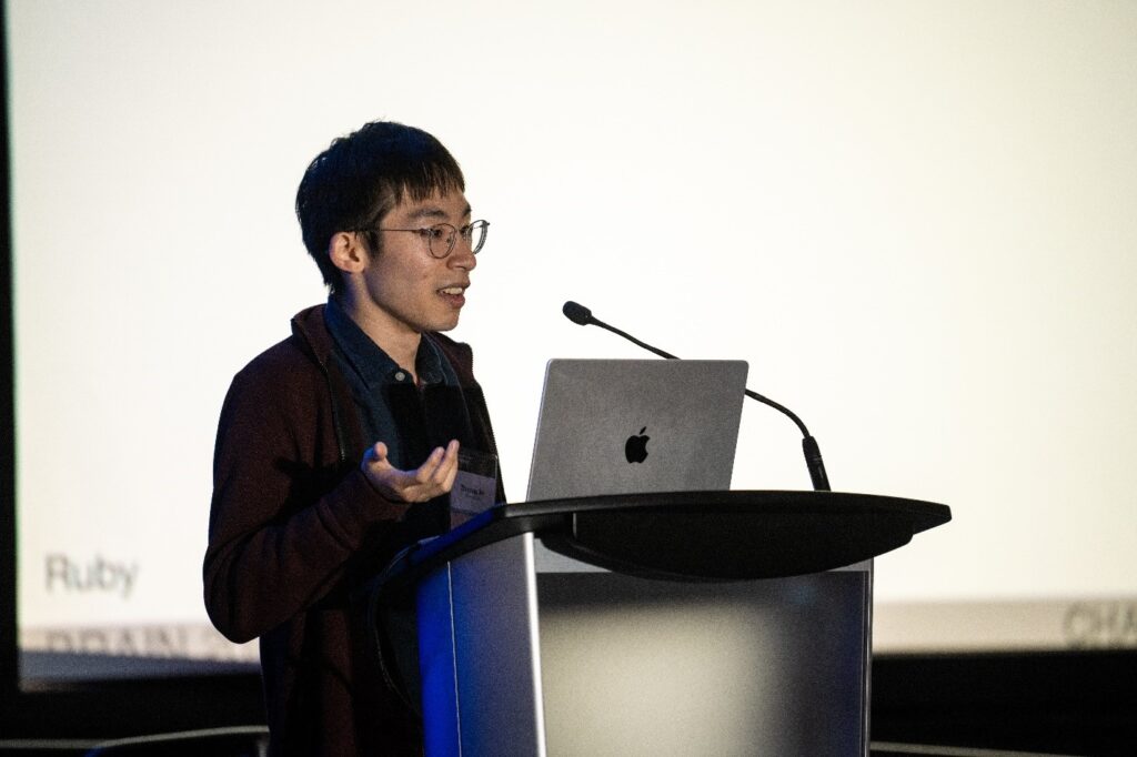
Figure 11. Dr. Zhiyang Jin presenting in the Imaging Outside-the-Toolbox: Novel Solutions for Large Scale and Deep-Tissue Imaging session.
Large-scale, functional neuroimaging is an evolving field, and we continue to see more new or upcoming solutions that experiment with chemical and genetic tools that address the limitations of current neuroimaging methods. Dr. Shy Shoham (New York University) moderated and gave the first talk in this session by explaining his optoacoustic tomography (OAT) efforts. OAT uses light pulses to generate ultrasonic waves and to produce an image, and his work uses near-infrared (NIR), genetically encoded calcium indicators to image calcium activity using optoacoustics. Dr. Takuya Terai (University of Tokyo) also discussed NIR calcium sensors and how his team uses them and chemigenetic fluorescent indicators for imaging. The team is working on new designs for indicators and exploring how to improve their performance.
Dr. Claire Deo (European Molecular Biology Laboratory) gave an overview of how her lab group develops chemigenetic labels and sensors for photoacoustic imaging. Her group uses this technology to label neurons in the mouse brain, aiming to figure out how to deliver fluorescent dye in vivo for whole brain labeling. Continuing the discussion on exploring brain activity patterns using calcium sensors, Dr. Hod Dana (Cleveland Clinic) discussed his lab’s methods for accessing brain regions of interest. To access larger brain areas, his team developed a new recording method using the CaMPARI (calcium-modulated photoactivatable ratiometric integrator) calcium integrator. Last, Dr. Zhiyang Jin (California Institute of Technology) presented on the advantages and prospects of ultrasound imaging readouts using molecular signals. Ultrasound calcium imaging is made possible using ultrasonic reporters of calcium (URoC) that can image receptor-specific calcium signaling in the mouse brain.
Symposium 3: Music: Building “Bridges” in the Brain
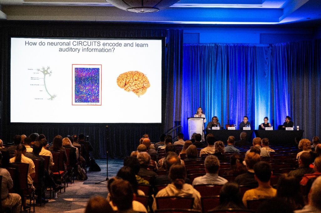
Figure 12. Dr. Maria Geffen speaking in the Music: Building “Bridges” in the Brain session.
In this session on music-based interventions, Dr. Emmeline Edwards (NIH National Center for Complementary and Integrative Health) opened by discussing the existing NIH efforts on music, neuroscience, and health. Dr. Maria Geffen (University of Pennsylvania) described her lab’s work in investigating the central auditory pathway to understand the mechanisms that neuronal circuits use to encode and learn sounds. Her team studies responses to frequent sounds as well as predictive signals, irregular sound detection, and memory formation through sound. Dr. Yuanyuan (Kevin) Liu discussed his work that explores if and how music or sound could decrease pain in mice. His research suggests that music can activate posterior and ventral posterior nuclei to help relieve pain if it has a low signal-to-noise ratio similar to ambient noise. “The mice really loved Bach, but not Mozart, or Beethoven,” joked Dr. Liu.
Next up, Dr. Malinda McPherson (Perdue University) discussed the underlying systems of pitch perception. Her research tested harmonic, inharmonic, and interleaved harmonic stimuli as well as delays to stimuli to see if they caused impaired memory performance. Her results suggested that people remember harmonic pitch better, making it critical for voice recognition but not for basic pitch discrimination. Dr. Narayan Sankaran (University of California, Berkeley) presented electrocorticography (ECoG) for neural encoding of melody to answer if the same neural populations encode pitch, pitch change, and expectations. Electrode findings indicated that while all computed from pitch, the properties of melody are encoded by different neural populations; however, the same electrodes encode pitch change in music and speech.
Symposium 4: Advancing Participant Engagement in Brain–Computer Interface (BCI) Research: Who, Why, and How?
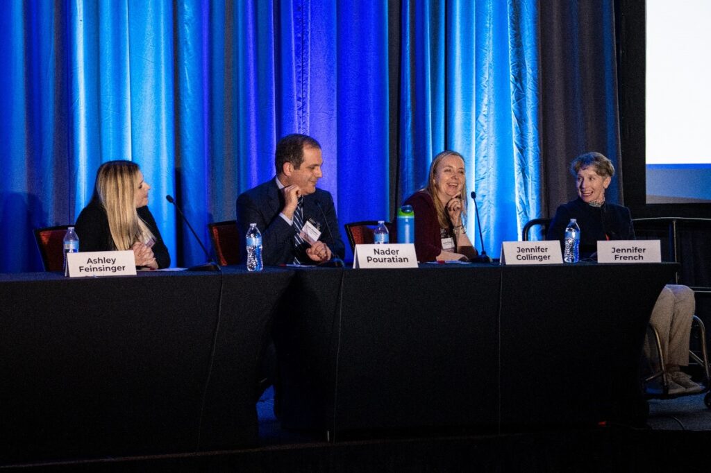
Figure 13. The panelists in the Advancing Participant Engagement in Brain–Computer Interface (BCI) Research: Who, Why, and How? session.
Dr. Ashley Feinsinger (University of California, Los Angeles) moderated this session and introduced brain–computer interface (BCI) research, focusing on implanted devices for real-time neural data processing. She also highlighted what participant engagement means in the scope of BCI research as well as the ethical, social, and technological opportunities and challenges associated with those types of studies. Jennifer French (Neurotech Network) spoke next, building on the introduction about participant engagement. She emphasized the importance of lived experience, explaining that after a snowboarding accident caused her paralysis, she joined a clinical trial for an implanted neuroprosthesis. Pulling from her own lived experience, she discussed the benefits and collaboration opportunities from engaging communities before, during, and after clinical trials.
Next, Dr. Jennifer Collinger (University of Pittsburgh) discussed the implanted BCI studies that she is involved with and the challenges she has seen in BCI participant engagement. She described a study design for an implanted sensorimotor device to help people with limb impairments that affect hand use and the lessons learned in future community engagement for studies like this. Dr. Collinger closed her presentation by introducing Phill McKenzie, a BCI user and study participant at the University of Pittsburgh. “I can’t move my arms or my legs, but I’m still trying to make a difference,” said Mr. McKenzie in a video that played after his introduction. Mr. McKenzie also joined the meeting remotely, telling the audience about his passion for music and how he made the decision to join an implant clinical trial. “I’m honored to be a part of this and to be with you all today,” he said. Last in this session, Dr. Nader Pouratian (University of Texas Southwestern) offered perspective from the lens of a physician and scientist, speaking about the BCI trials he has been involved with and his goals for participant engagement. He closed by discussing challenges for engagement, such as the trial and error involved, the lack of a formal system on how to engage patients, and how to make clinical trials more adaptive based off participant feedback.
Symposium 5: Building on the BRAIN Initiative Cell Census Network (BICCN): Alzheimer’s Disease Multimodal Atlasing Projects (AD-MAPs)
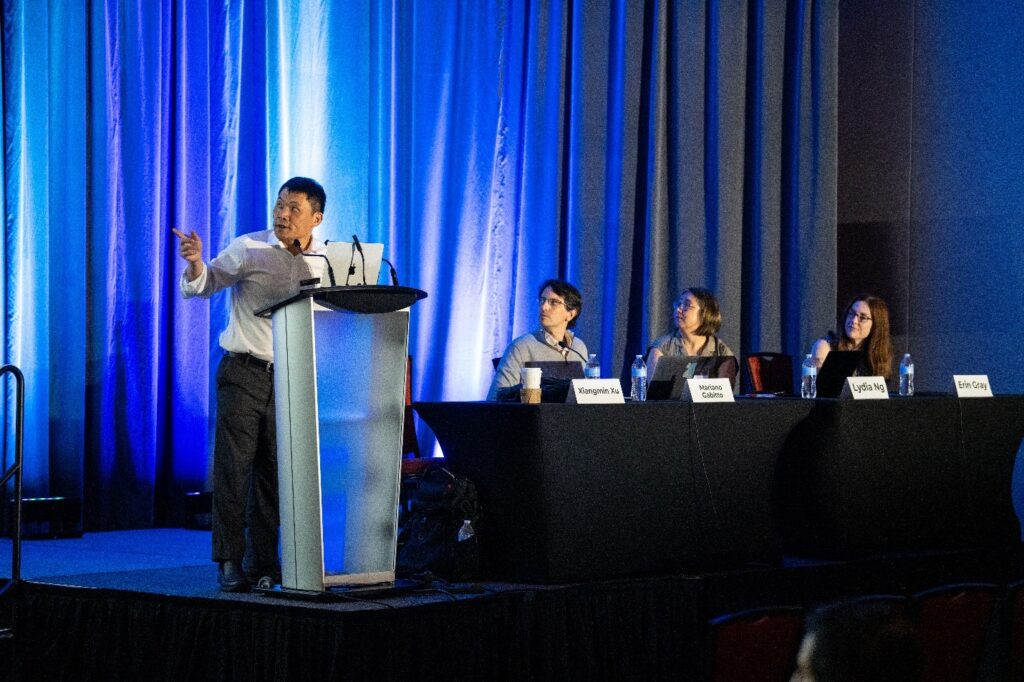
Figure 14. Dr. Xiangmin Xu at the podium of the BICCN: AD-MAPs session.
This session, organized by Dr. Erin Gray of the National Institute on Aging (NIA), was opened by moderator Dr. Xiangmin Xu (University of California, Irvine). Dr. Xu discussed AD-MAPs, explaining that the NIA’s goal is to build on BICCN resources to create high-resolution, multimodal atlases that help understand circuit changes related to aging and Alzheimer’s disease. A prerecorded video featuring NIA Director Dr. Richard Hodes further iterated the importance of the BICCN for forming the AD-MAPs. Dr. Xu returned to give his talk on hippocampal circuit mapping and single cell, spatial transcriptomics in Alzheimer’s disease mouse models. Dr. Xu’s research hopes to better understand brain circuit connectomes to diagnose and treat Alzheimer’s disease earlier and better. The next talk featured Dr. Maura Boldrini (Columbia University), who presented data that combined spatial transcriptomics and single nuclei ribonucleic acid (RNA) and assay for transposase-accessible chromatin (ATAC) sequencing to identify individually expressed genes in major depressive disorder (MDD). She discussed strategies her team uses to identify cell clusters in the human hippocampus and to identify neurogenetic niche cell types related to MDD.
Dr. Mariano Gabitto (Allen Institute for Brain Science) followed by presenting on cellular analysis use cases for the Seattle Alzheimer’s Disease Brain Cell Atlas (SEA-AD) to learn more about cell types and molecular targets related to Alzheimer’s disease and to eventually understand how the disease progresses in the brain. Complementary to Dr. Gabitto’s talk, Dr. Lydia Ng (Allen Institute) closed the session by discussing the data available in the SEA-AD and how it is made available for the wider research community. Dr. Ng discussed what researchers can do with the tools in SEA-AD, including how to map cells to BICCN/BICAN references and how to filter donor data.
Symposium 6: BRAIN Informatics in the AI Era
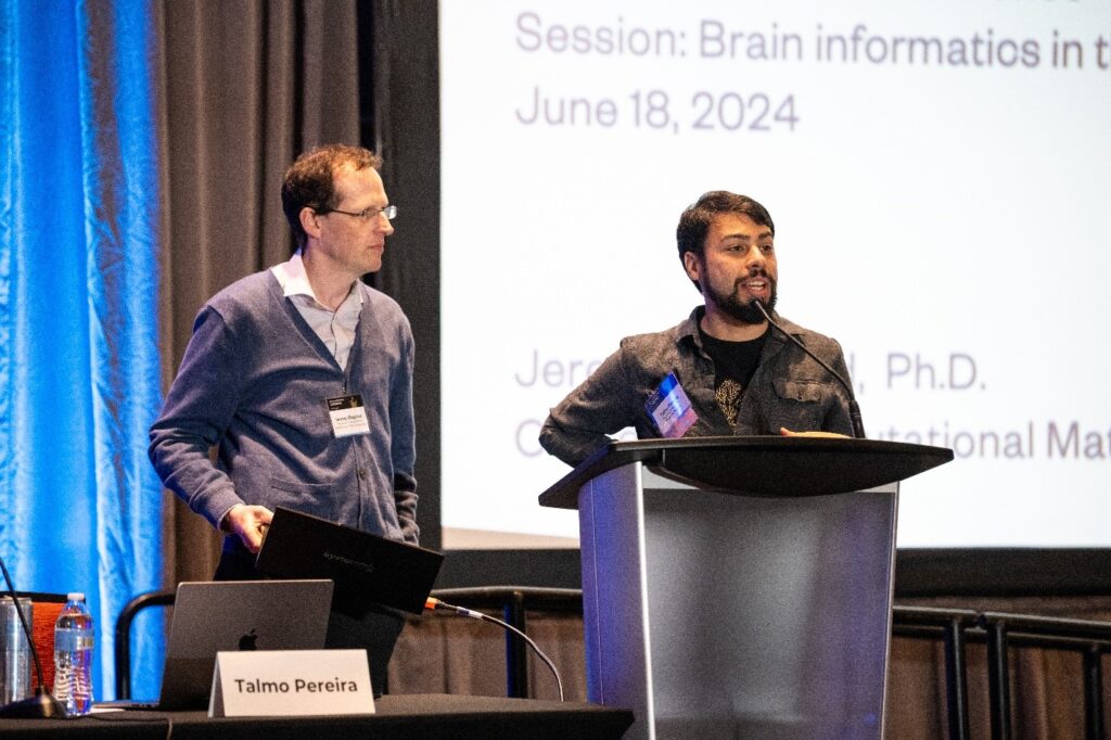
Figure 15. Dr. Talmo Pereira (right) introducing Dr. Jeremy Magland (left) in the BRAIN Informatics in the AI Era session.
Dr. Talmo Pereira (Salk Institute for Biological Studies) moderated this session on informatics and AI, stating that great computational power comes with great challenges. Liezl Maree (Salk Institute for Biological Studies), a member of Dr. Pereira’s lab, presented the lab’s work developing Social LEAP Estimates Animal Poses (SLEAP), a cloud-based tutorial platform. SLEAP uses computer vision and deep learning to go markerless and allow scientists to capture all forms of biological motion. Its move to the cloud is recent but has overcome accessibility roadblocks such as graphic processing units (GPU) and desktop software installation.
Next, Dr. Jeremy Magland (Flatiron Institute) showcased three browser-based tools for data visualization and analysis. Figurl is a visualization tool that creates shareable interactive figures from complex data using a clickable hyperlink method for browser use. Neurosift is another visualization tool that explores the Distributed Archives for Neurophysiology Data Integration (DANDI) Archive and views Neurodata Without Borders (NWB) files locally and remotely. Dendro is an early-stage automated data analysis web application that will process DANDI data sets in the cloud. Last, Dr. Satrajit Ghosh (Massachusetts Institute of Technology) spoke on the AI infrastructure that enables these kinds of neuroscience tools to be developed. He said that data standardization is critical so that an AI system can understand what happens to data over time and so that data sets are accessible through a programmable interface. With these standardizations come many challenges related to data scalability and modality, as well as AI’s ability to understand data sets living in different ecosystems that do not always interact with each other. Dr. Ghosh hopes that in the future, data will continue to rely on FAIR principles (findable, accessible, interoperable, reusable) to create more seamless AI infrastructure.
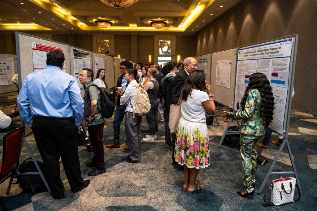
Poster Sessions and Exhibits
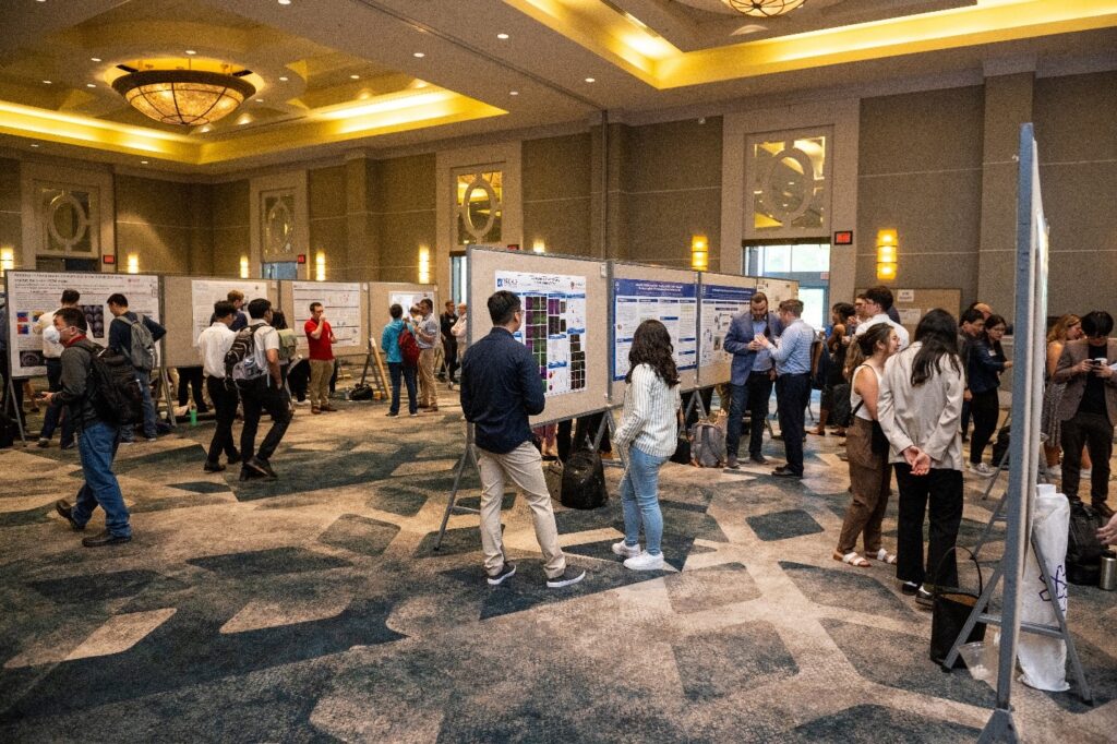
Figure 16. The poster hall at the 10th Annual BRAIN Initiative Conference.
Another highlight of the 10th Annual BRAIN Initiative Conference was the large poster hall lined with presenters showcasing the latest installments in their research. During the three poster sessions, attendees flocked to the hall to listen to the poster presenters and to ask questions about their work. If you registered for the conference, you can login using your event credentials to view and download the posters on the conference website, where you can toggle by topic to explore any posters you may have missed.
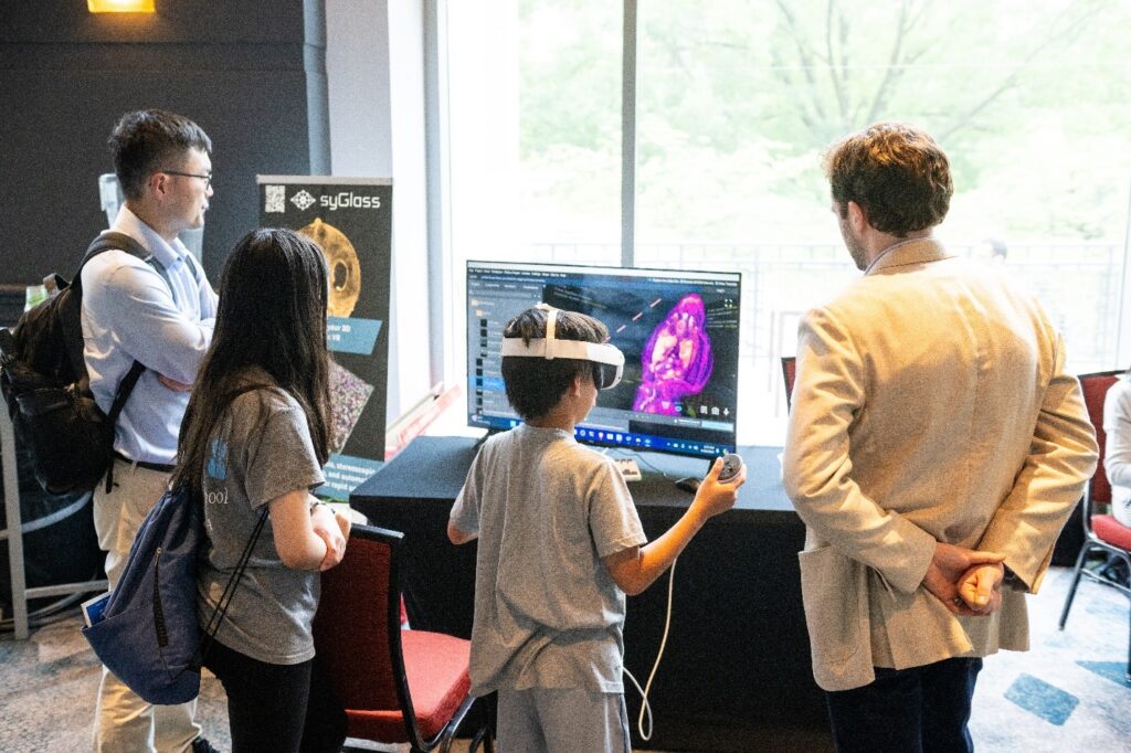
Figure 17. Children experimenting with syGlass’s virtual reality, with Lead Software Engineer Nathan Spencer on the far right.
In addition to the poster hall, exhibitor booths featured programs, resources, and interactive tools. For example, syGlass (pictured above) is a software system for 3D and 4D image visualization and virtual reality. The syGlass team is starting to connect researchers to high school students with an interest in learning more about neuroscience earlier in their careers. “Moving into education and high school classrooms will bridge the gap between research and education,” said Nathan Spencer, syGlass’s lead software engineer.
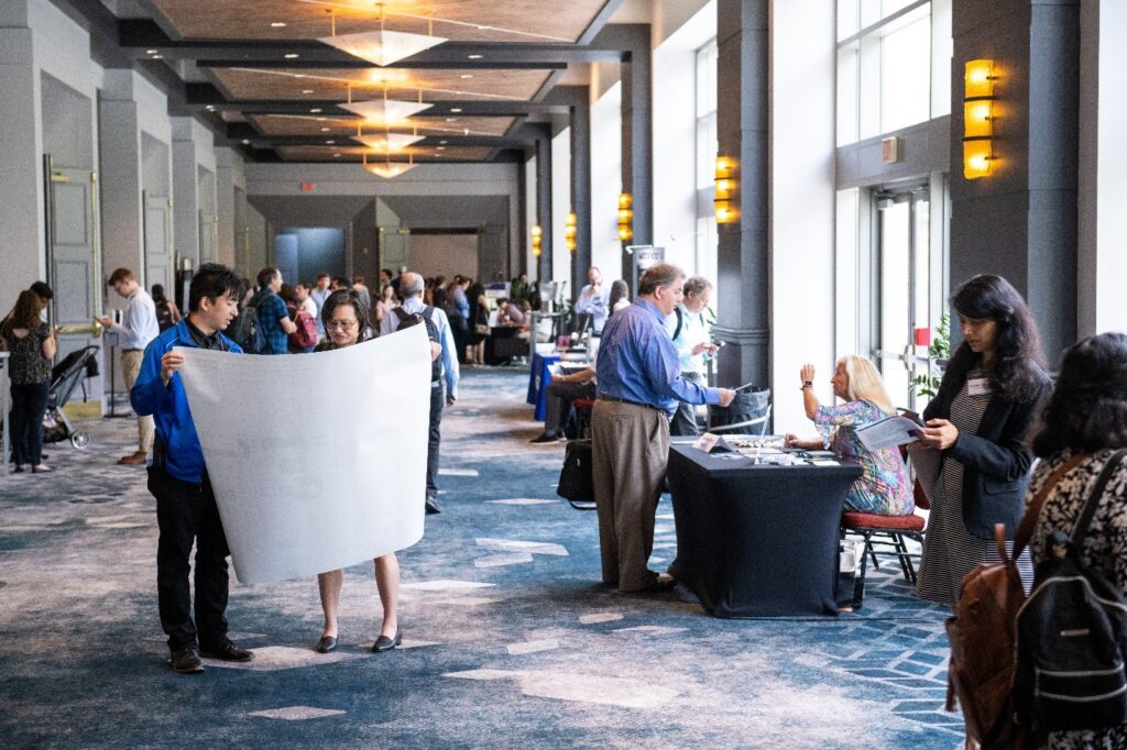
Figure 18. The main hallway of the 10th Annual BRAIN Initiative Conference lined with exhibitor booths.
The Brain Donor Project is another example of an exhibitor featured at the conference. This nonprofit supports the brain banks at the NIH, helping people arrange to become brain donors when they die. Tish Hevel, CEO and founder of The Brain Donor Project, said, “The best part of being here is talking to people who are excited about what they’re doing to make us more neurologically advanced.”
Specialty Sessions
BRAIN, Neuroscience, and Beyond: Building Our Early Career Community
Figure 19. Early career researchers and Dr. Ngai mingling at the early career community event.
Early career researchers were invited to attend an event the day before the conference began to network, learn about funding opportunities, and meet staff from the BRAIN Initiative. “I really enjoyed the early career community event the day before the conference. There were so many investigators to share ideas with, it was a great time,” said Dr. Zheng Zhang (Yale University).
BRAIN and the Future of Computing: Emerging Perspectives on Embodied NeuroAI Research
AI was a major theme at the conference, including during this session focusing on NeuroAI, or the combined power of neuroscience, behavior, and AI. Dr. Tony Zador (Cold Spring Harbor Laboratory) introduced NeuroAI and moderated a panel, including Dr. Gina Adam (The George Washington University), Dr. Kim Stachenfeld (Google DeepMind and Columbia University), and Dr. Henry Yin (Duke University). Panelists spoke about the implications of neural control using computing, graph neural network approaches, and biomedical applications of neurotechnologies.
Bringing Your Neurotechnology to Life: Topics on Translation
In this workshop, panelists Jennifer French (Neurotech Network), Dr. April Marrone (U.S. Food and Drug Administration), Dr. Evaristus Nwulia (Howard University), Katie Sale (American Brain Coalition), and Dr. Vanessa Tolosa (Meta) discussed funding strategies and resources for neurotechnology, including the NIH Blueprint’s NeuroTech Course.
Fostering Inspiration and Engagement through Outreach, Noggins and Art
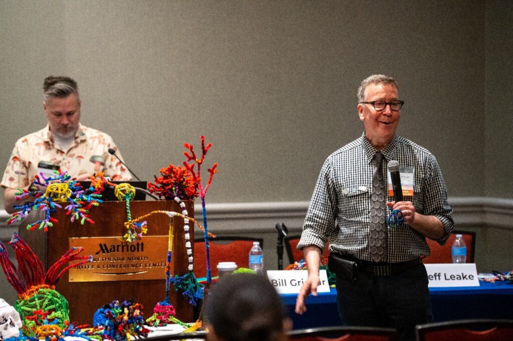
Figure 20. Dr. Bill Griesar and Jeff Leak opening their session on neuroscience and art.
In this in-person interactive session, participants embraced their creative side by examining three-dimensional brain models and building brain cells using colorful pipe cleaners. Dr. Bill Griesar and Jeff Leake (Portland State University) are part of a nonprofit called NW Noggin, which organizes interactive activities and workshops like this to introduce and excite today’s youth about neuroscience using art.
Technology and Resource Dissemination: Best Practices and Lessons Learned
This in-person panel session was moderated by Dr. Natalie Trzcinski (National Institute of Neurological Disorders and Stroke) and featured Dr. Charla Howard (Spike Neuro), Dr. Christopher Hoy (Meadowlark Optics), Dr. Mattias Karlson (SpikeGadgets), and Dr. Shaheen Latif (Pinnacle Technology). The panel opened by discussing barriers to commercialization and lessons learned in tool engineering. Later, the panelists spoke on their transitions from academia to small business, told stories about how they knew when to let go of certain technologies, and discussed how to determine if there is a need for a technology and how they approach licensing, patents, and product sustainability.
Brain Behavior Quantification and Synchronization (BBQS) – Laying the Foundation for a New Data and Knowledge Ecosystem
The BRAIN Initiative’s BBQS program was created to develop new tools and ways to understand the mechanisms of behavior. The program’s consortium kicked off its activities during this session for current and future BBQS investigators, opening with Dr. Sarah Hollingsworth (Holly) Lisanby (National Institute of Mental Health) introducing the program’s goals before Dr. Satrajit Ghosh (Massachusetts Institute of Technology) presented on how machine learning and AI will be used in the program. Dr. Ashley Feinsinger (University of California, Los Angeles) covered BBQS project neuroethics, and Dr. Maryam Shanechi (University of Southern California) discussed the engineering and mathematical approaches for BBQS data.
Meet the Funders Networking Session
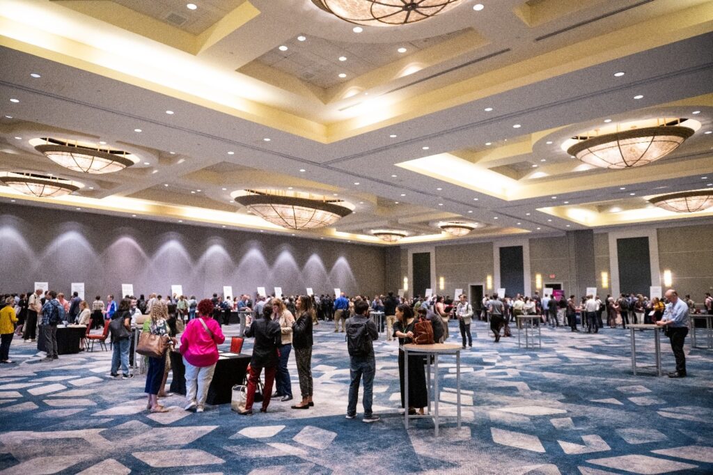
Figure 21. At the Meet the Funders session, participants stopped by tables from partner organizations to network.
This networking opportunity allowed attendees to gather and mingle with BRAIN Initiative partner organizations to learn more about funding opportunities. This session had representation from the NIH BRAIN Initiative, Simons Foundation, National Science Foundation, Dana Foundation, and more. Virtual attendees could also submit questions for the funders to respond virtually at a later time.
Conclusion
Dr. Ngai closed out the conference by thanking everyone for attending and for their contributions to brain science over the past 10 years. Throughout the conference, attendees shared what they thought the next decade of brain research could look like:
- “There are a lot of neuroscience tools and methods that are both in the infancy stage and some that have already been studied for a very long time. I think there’s a lot of room for growth in the next 10 years to utilize new ways to study the brain.” —Ciara Bagnall-Moreau (The Feinstein Institutes for Medical Research)
- “In the next 10 years, I’d love to see more BRAIN Initiative early career grants—I look forward to having opportunities to apply.” —Taruna Yadav (Yale University)
- “In 10 years, I’d love to see us talking about not just laying down the foundation or treatments, but cures and preventions based off all the great science that we’re doing.” —Dr. John Ngai
We are grateful for everyone who helped us celebrate a decade of innovation at the 10th Annual BRAIN Initiative Conference!
All images provided by Lesnick Photo.