Summary: NIH’s BRAIN Update Blog has published its latest roundup of recent BRAIN Initiative-funded research papers.
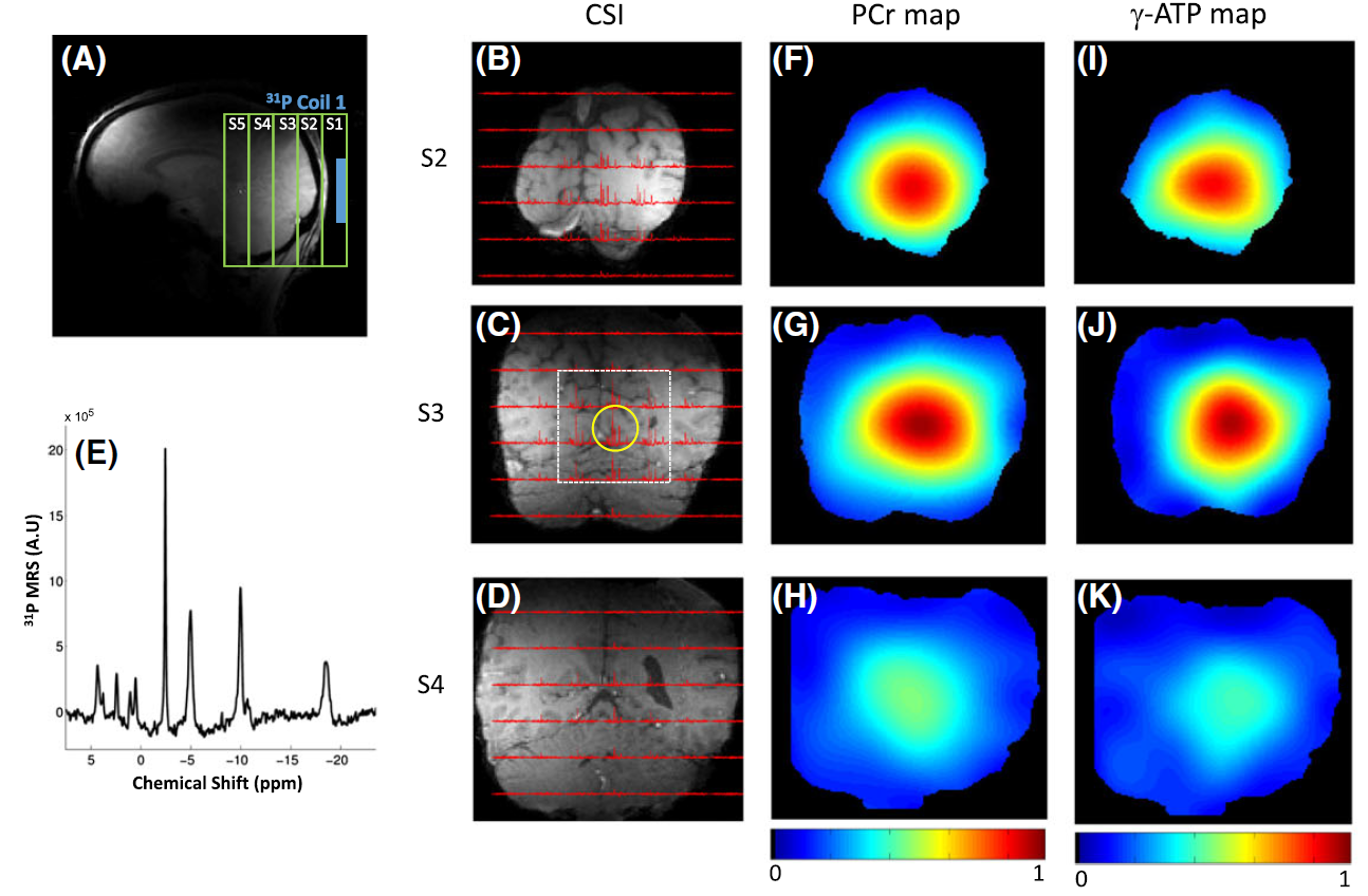
Representative in vivo phosphorus‐31 three‐dimensional chemical shift imaging (CSI) data detected in the human occipital lobe, as well as maps of phosphorus metabolite signals (phosphocreatine (PCr) and γ‐adenosine triphosphate (γ‐ATP)) overlaid on the anatomical images. (A) Location of the coil placed near the occipital lobe. (B–D) Phosphorus-31 MR spectroscopy (MRS) profiles from three selected CSI slices. (E) Single-voxel phosphorus-31 spectrum denoted by the yellow circle in (C) shows excellent detection sensitivity. (F–H) Corresponding PCr maps. (I–K) Corresponding γ-ATP maps. Similar images were obtained simultaneously from the frontal lobe.