Summary: The entire fruit fly brain has been imaged by scientists at Howard Hughes Medical Institute, a partner institution of the BRAIN Initiative.
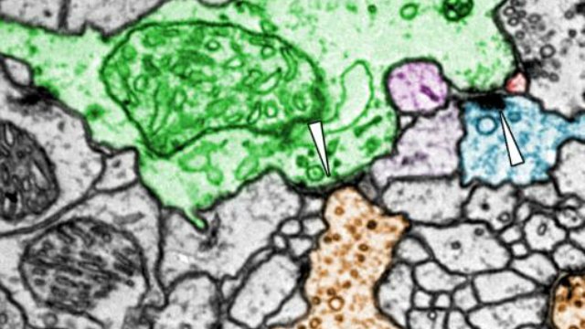
A database of electron microscopy images reveals the connections of the entire female fruit fly brain. In this image, types of Kenyon cells (KC) in the mushroom body main calyx are labeled by color: αβc-KCs are green, αβs-KCs are yellowish brown, and gamma-KCs are blue. The white arrows point to visible presynaptic release sites.ZHENG ET AL. 2017