In 2020, BRAIN researchers continued to create new, state-of-the-art technology. We also witnessed advances in open science and data availability – movements that benefit of the entire scientific community. Here, we will look back on the year 2020, as well as look ahead to what the world of neuroscience is bringing in 2021.
Looking Back…
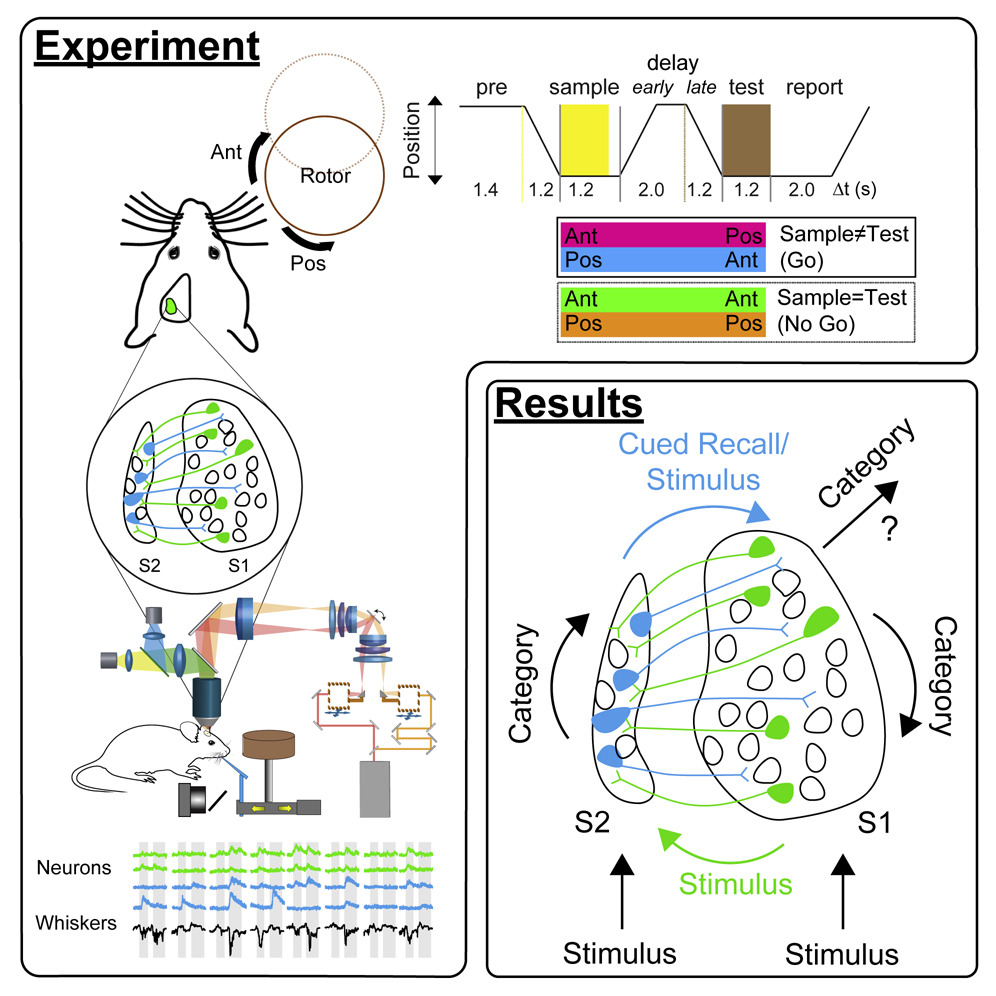
Could a memory game help us understand brain injury? This year, NSF-funded BRAIN researchers used optical imaging and optogenetic technology to dissect the role of the primary and secondary somatosensory cortices (S1 and S2, depicted above) to pinpoint which skills may be harder to recover following traumatic brain injury. They used a whisker memory game in mice to reveal that specific types of task-related information occur in both segregated and distributed manners across both brain regions. National Science Foundation News also highlighted this work, which could lead to a better understanding of how the human brain handles sensory information and relearns motor skills.
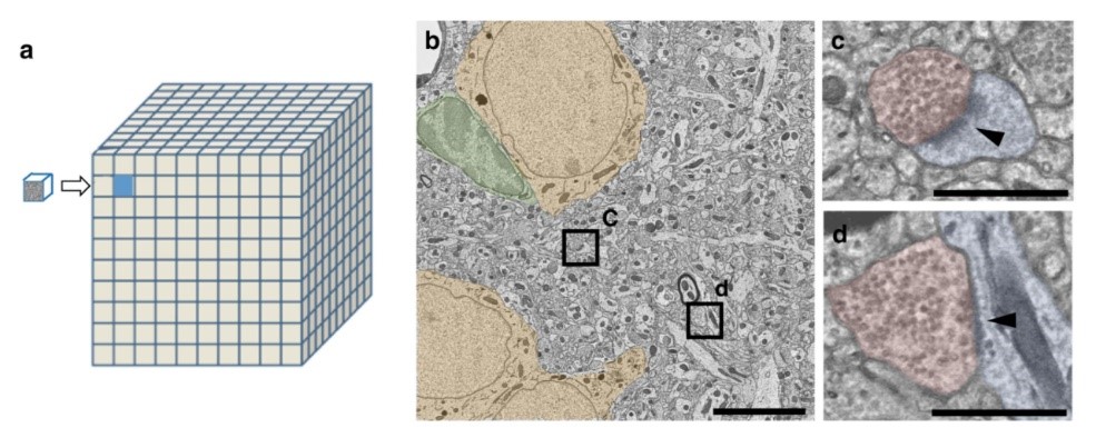
The IARPA MICrONS program also was a rousing success in the year 2020. For instance, IARPA-funded BRAIN researchers developed a petascale automated imaging pipeline for mapping neuronal circuits using high-throughput transmission electron microscopy. This work demonstrates the feasibility of acquiring large electron microscopy datasets at the level of cortical microcircuits across multiple brain regions. The image above shows a cubic millimeter of mouse cortex (a) as visualized using an electron microscope (b). Another advantage of this development in imaging is that it can be implemented in large research centers, as well as distributed over a number of smaller, individual laboratories.
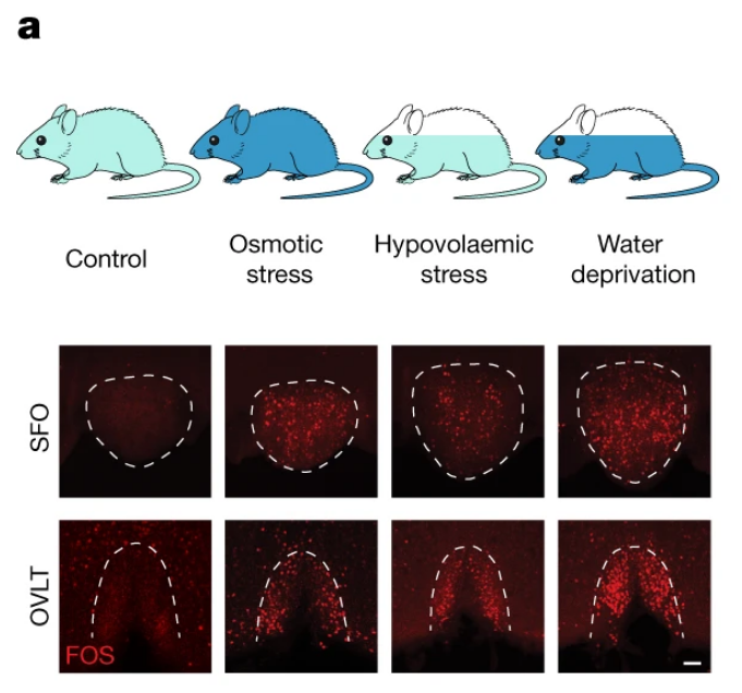
How does the brain quench thirst? One group of NIH-funded BRAIN researchers made quite the splash this year when they examined the cellular basis of different types of thirst in rodents. The two types of thirst they examined were osmotic and hypovolaemic thirst. Osmotic thirst is when we crave pure water, whereas hypovolaemic thirst is when we crave water with minerals. In the image above, elevations of blood osmolality, as well as the loss of body fluid induces robust c-Fos (an indirect marker of neural activity) expression in both the subfornical organ (SFO) and the organum vasculosum of the lamina terminalis (OVLT). Last year, NPR covered these findings in an article, “Water Or A Sports Drink? These Brain Cells May Decide Which One We Crave.” This work would not have been feasible without the open and welcoming environment espoused by U.S. universities in general and Caltech in particular,” said one researcher involved with this study in an article by Caltech News, thereby highlighting the importance of open borders in science.
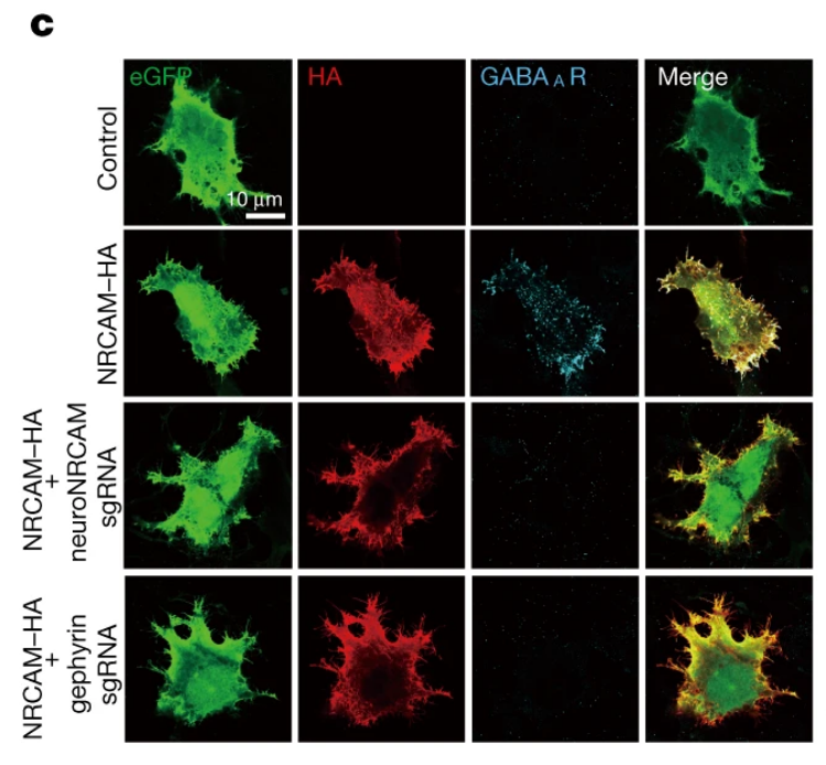
Astrocytes are the “master conductors” of the brain. This was, more or the less, the running headline across several science news outlets last year, including Neuroscience News, The Life Boat Foundation, and NR Times. This NIH-funded discovery published in 2020 was truly revolutionary. By using sophisticated viral and genetic techniques, these BRAIN researchers demonstrated that an astrocytic adhesion molecule called NRCAM regulates inhibitory, but not excitatory, synapses within the brain. “We really discovered that the astrocytes are the conductors that orchestrate the notes that make up the music of the brain,” said senior author and BRAIN researcher Dr. Scott Soderling in an article by Duke University News.
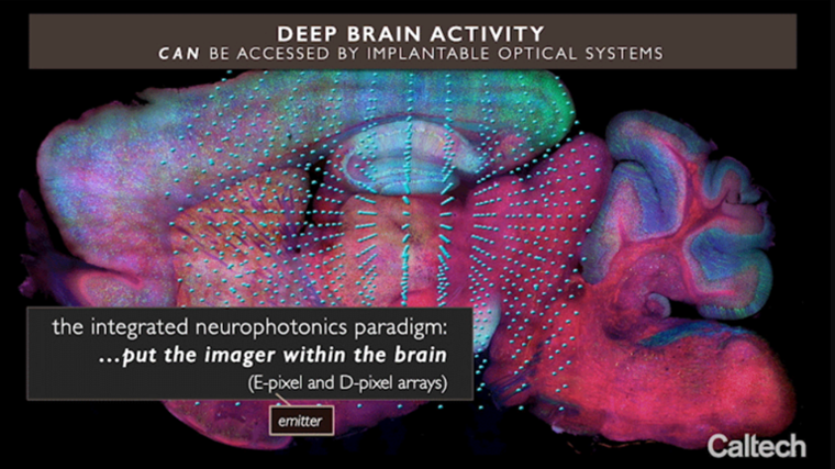
The year 2020 also fostered studies that spanned multiple BRAIN Initiative Alliance organizations. For instance, BRAIN Initiative funding allowed NIH, NSF, the Kavli Foundation, and DARPA to support an international collaboration aimed to realize “integrated neurophotonics,” defined by the research team as a combination of the advances in microchip-based integrated optical and electronic circuitry with advances in optogenetics. In their 2020 paper, these BRAIN researchers demonstrated the potential of their integrated neurophotonics paradigm in achieving fast, dense, and deep functional imaging in brain tissue. This video (image still above) depicts the integrated neurophotonics process. BRAIN researcher Dr. Michael Roukes told Caltech News, “Dense recording at depth—that is the key. We will not be able to record all of the activity of the brain any time soon. But could we focus on some of its important computational structures within specific brain regions? That’s our motivation.” This study was also featured in Psychology Today.
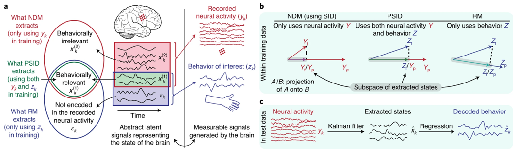
Can researchers isolate and decode brain signal patterns for specific behaviors? In 2020, a team of NIH- and NSF-funded BRAIN researchers sought to answer this very question. These BRAIN researchers developed a machine-learning algorithm called preferential subspace identification (PSID) that resolves several of the problems associated with isolating neural patterns related to specific behaviors. Dr. Maryam Shanechi told USC Viterbi News, “We have developed an algorithm that, for the first time, can dissociate the dynamic patterns in brain signals that relate to specific behaviors one is interested in.” Algorithms such as this have the potential to help researchers develop enhanced brain-machine interfaces to restore lost brain function in those with neurological and mental disorders.
2020 Researcher Spotlight…

Dr. Lucas Cheadle, from Cold Spring Harbor Laboratory, was named one of the Allen Institute’s 2020 Next Generation Leaders. As a Next Generation Leader, Cheadle will serve as an advisory board member for the Allen Institute for Brain Science and the MindScope Program. Additionally, Cheadle is the first NIH BRAIN K99 grantee to transition to the R00 phase of his award. His BRAIN Initiative project is aimed at understanding a novel microglia-to-neuron signaling pathway. Congratulations Dr. Cheadle!
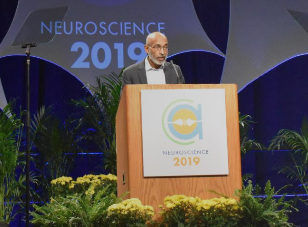
Of course, there are many other exceptional BRAIN researchers worthy of recognition this year. For instance, Drs. Aviv Regev and Edward Chang were elected to become members of the National Academy of Medicine. Dr. Emery Brown (pictured above) won the Society for Neuroscience’s Swartz Prize for Theoretical and Computation Neuroscience. Dr. György Buzsáki won the Society for Neuroscience’s Ralph W. Gerard Prize in Neuroscience. Additionally, Dr. Robert Desimone won a Brain and Behavioral Research Foundation Goldman-Rakic Prize for Outstanding Achievement in Cognitive Neuroscience Research. Drs. Jerold Chun, Duygu Kuzum, and Christopher D. Harvey were recipients of 2020 NIH Director’s Awards. Finally, spatially resolved transcriptomics, a method developed by Drs. Hongkui Zeng, Xiaowei Zhuang, and other colleagues at the BRAIN Initiative Cell Census Network (BICCN) project, was crowned Nature’s “Method of the Year 2020.”
Looking Ahead…
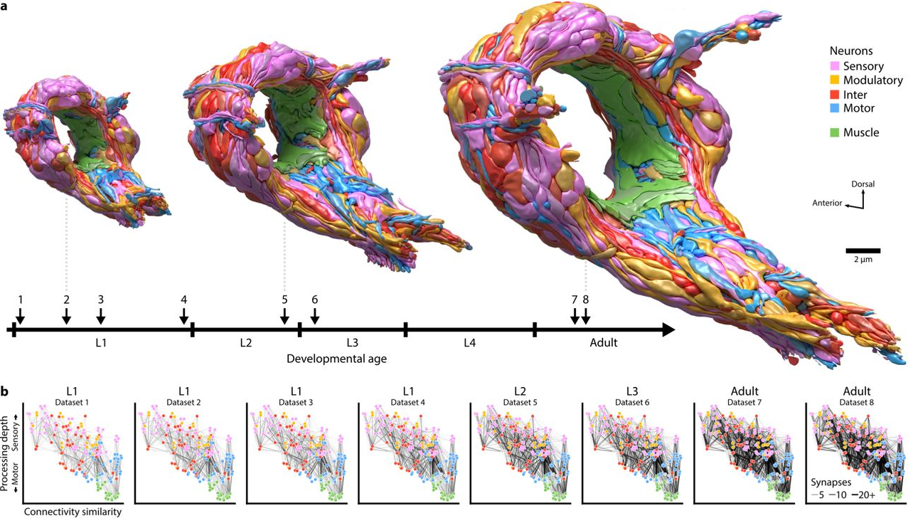
If these 2020 preprints are any indication, the year 2021 is likely to see myriad innovative publications. For instance, NSF-funded BRAIN researchers are mapping brain maturation across development in C. elegans. The image above is a depiction of the developmental timeline of eight reconstructed C. elegans brains, with topological models shown at three stages.
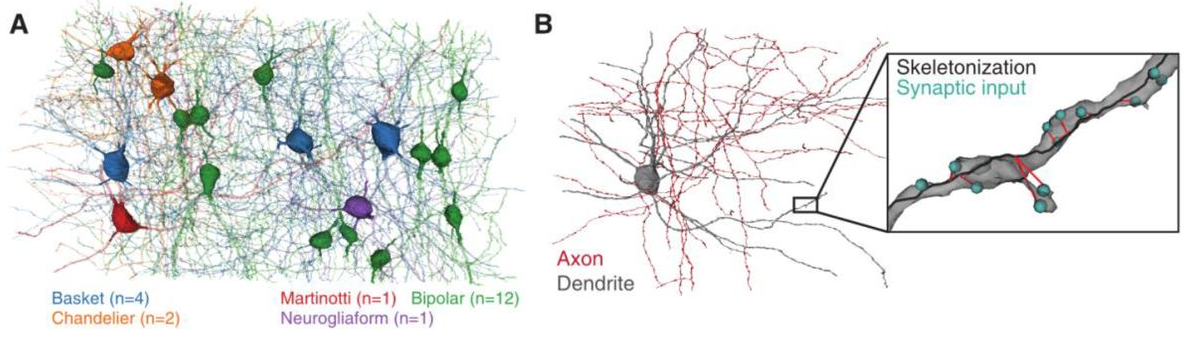
Another promising preprint comes from the IARPA-funded BRAIN researchers supporting the MICrONS project. These BRAIN researchers are using electron microscopy to establish multiscale and multimodal reconstructions of cortical structure and function (inhibitory interneurons are shown in (a) and an example basket cell are shown in (b)). Many of these data and analysis pipelines are openly available. Electron microscopy imaging data and cellular segmentation can be viewed at: https://microns-explorer.org/. The raw calcium imaging data are available together with the analysis code at: https://github.com/seung-lab/MicronsBinder. Further, the code for generating the cell and organelle reconstructions is available here: https://github.com/seung-lab/. Altogether this study represents another important leap forward in open science and data availability.
Looking back, the achievements of the U.S. BRAIN Initiative and BRAIN researchers were astounding in 2020. However, there is sure to be more to come in the year 2021! Be sure to mark your calendar (or download the new 2021 BRAIN Initiative calendar) for the 7th Annual BRAIN Initiative Investigators Meeting. This virtual event will be held June 15-17, 2021. If it’s anything like the 2020 Virtual Investigator’s Meeting, it’s bound to be a big success! Finally, check out our new BRAIN Initiative Alliance Toolmakers’ Resources page and Toolmakers Newsletter! Without a doubt, 2021 will have much in store for neuroscience, neurotechnology, and the U.S. BRAIN Initiative.